Usage
Fill in the phantom with a dilution containing 2.7 MBq of F18-FDG. This is an approximate value; small deviations are not relevant. Higher activities are also possible, but they will lead to an unnecessary dose exposure and longer processing time to generate images.
For further information concerning the positioning, image settings and acquisition parameters refer to the Accompanying Documentation of your PET/MR-system, or contact the Bruker Service.
Please make sure you have read the safety section on this manual first.
Prior to filling the phantom, take precautions for liquid spills, e. g. paper towels.
Personal protective equipment goggles and gloves are required. Contaminated clothes must be changed immediately. Check your gloved hands for contamination after filling the phantom.
The suggested procedure for filling the phantom is:
- 1.
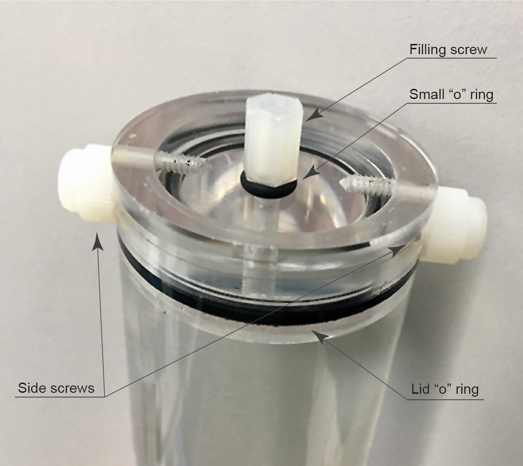
- Remove top plastic screw. It is assumed that the top lid with its large “o” ring seal is fitted with the 2 side screws.
- 2.
- Fill the phantom in with distilled water to 90% of its capacity. Preferably use a large (20-70 ml) syringe.
- 3.
- Add the radioactive solution with a suitably sized syringe (1-2 ml) and long needle.
- 4.
- Top up with distilled water. Use the filling hole as an air bubble trap. Minimize the size of the air bubble by carefully sucking out the air.
- 5.
- Close with the top plastic screw making sure its associated “o” ring seal is present.
- 6.
- Wipe out the outside of the phantom to make sure there are no spills on the surface.
- 7.
- Shake gently.
- 8.
- Place the phantom without its holder inside the dose calibrator and note the activity and time of the reading. When taking activity readings, make sure the phantom is fully inside the calibrator well. The dose calibrator needs periodic calibrations to ensure the readings are reliable.
Close-up view of the NMI PHAN IM QA D30
The recommended acquisition settings are:
- ▪
- Acquisition duration: 1200 s
- ▪
- Reconstruction settings: MLEM, 0.75 mm voxel and 12 iterations.
A typical pitfall in these acquisition is not to have the phantom axially centered. It is advisable to carry out a localizer study for 10 s and 3 iterations to find out the correct axial location.
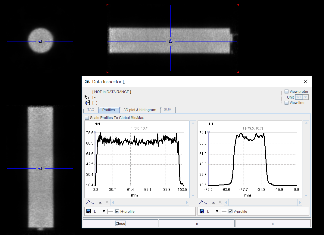
After imaging on the PET system, they produce an image which matches the size of the fillable volume inside the phantom. In normal operating conditions the image will be uniform.
Coronal/Sagittal and transverse PET Images of the NMI PHAN IM QA D30 3R


