Setup
Animal Position and Fixation
The animal can be positioned in the cassette head first (respiration mask according to the first figure below) or tail first (respiration mask according to the second figure below). Depending on the animal size, additional fixation of the animal can be done using tape to avoid displacement of the animal during measurement or transfer of the cassette from one imaging modality to another.
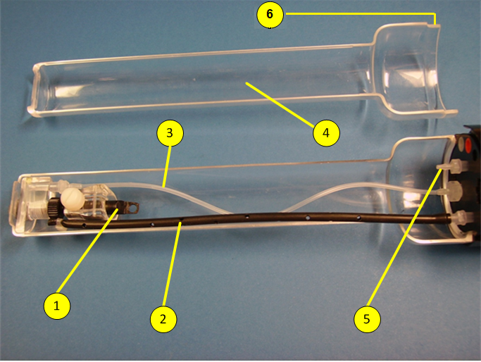
Small Mouse Cassette: Tooth bar and respiration mask mounted in head first position. In this setup, the anesthesia gas is supplied to the nose cone via the silicon tube (3).
Respiration Mask, Anesthetic Gas Supply and Extraction
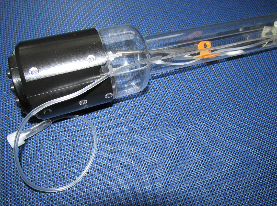
The respiration mask in head first position (see figure above) is supported via a silicon tube (3) to supply the anesthesia gas through the tooth bar. In tail first position, the silicon tube is removed, the tooth, bar and the respiration mask are unscrewed and attached on the opposite site of the cassette where initially the silicon tube was located (see figure below).
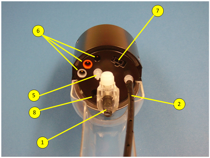
In both setup, the anesthesia gas exits the nose cone of the respiration mask and distributes into the cassette. From there, is extracted through suction hole (8).
Small Mouse Cassette Interface: Tooth bar and respiration mask mounted in tail first position. In this setup, the anesthesia gas is supplied to the nose cone directly through the interface.
Animal Respiration, Temperature Control and ECG
The cassette has connectors for respiratory monitoring (5), ECG (6), and temperature monitoring (7). The connectors are compatible with the monitoring system of SA Instruments Inc. (www.i4sa.com).
Warm Air Supply
Warm air is supplied via a diffusor tube (2) positioned along the cassette to stabilize the body temperature of the animals during investigation after the cassette cover (4) is closed.
Depending on the modality used, the air supply has to be connected to the corresponding heating system.
Catheter Port
Use the opening at the end of the cassette cover as port for a catheter hose.


