 “Assessment of a 1H high-resolution magic angle spinning NMR spectroscopy procedure for free sugars quantification in intact plant tissue” Teresa Delgado-Goñi, Sonia Campo, Juana Martín-Sitjar, Miquel E. Cabañas, Blanca San Segundo, Carles Arús; Planta, 238:397-413, 2013 (PDF file). DOI: 10.1007/s00425-013-1924-y
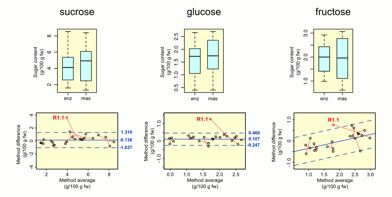
In most plants, sucrose is the primary product of photosynthesis, the transport form of assimilated carbon, and also one of the main factors determining sweetness in fresh fruits. Traditional methods for sugar quantification (mainly sucrose, glucose and fructose) require obtaining crude plant extracts, which sometimes involve substantial sample manipulation, making the process time-consuming and increasing the risk of sample degradation. Here, we describe and validate a fast method to determine sugar content in intact plant tissue by using high-resolution magic angle spinning nuclear magnetic resonance spectroscopy (HR-MAS NMR). The HR-MAS NMR method was used for quantifying sucrose, glucose and fructose in mesocarp tissues from melon fruits (Cucumis melo var. reticulatus and Cucumis melo var. cantalupensis). The resulting sugar content varied among individual melons, ranging from 1.4 to 7.3 g of sucrose, 0.4–2.5 g of glucose; and 0.73–2.83 g of fructose (values per 100 g fw). These values were in agreement with those described in the literature for melon fruit tissue, and no significant differences were found when comparing them with those obtained using the traditional, enzymatic procedure, on melon tissue extracts. The HR-MAS NMR method offers a fast (usually <30 min) and sensitive method for sugar quantification in intact plant tissues, it requires a small amount of tissue (typically 50 mg fw) and avoids the interferences and risks associated with obtaining plant extracts. Furthermore, this method might also allow the quantification of additional metabolites detectable in the plant tissue NMR spectrum.