Workshop limited to 4 participants (first come, first served)
Contact person:
Silvia Lope-Piedrafita, PhD ()
This course combines a comprehensive series of lectures on the technology of Magnetic resonance spectroscopy and imaging (MRS/MRI) with hands-on laboratory sessions to provide practical demonstrations of key concepts and procedures for preclinical studies.
Whether you are considering MRI as a research tool in your lab or just would like to learn more about MRI, this workshop addresses practical aspects of experimental MRI with laboratory animals and provide valuable hands-on experience on a 7 Tesla Bruker BioSpec spectrometer.
Last 26th July 2023 I successfully defended my PhD Thesis entitled: “PhD Thesis: Implementation of high-resolution MRSI methods in a pre-clinical scanner, and optimization for brain longitudinal studies of therapy response in mice glioma model.”
Abstract
Magnetic resonance spectroscopy and magnetic resonance spectroscopic imaging (MRS/MRSI) are non-invasive diagnostic techniques that use a strong magnetic field and radio waves to examine the chemical composition of living tissue. Working on the same principles as Magnetic Resonance Imaging (MRI), instead of producing images, MRS generates a spectrum of signals that can be used to identify the type and amount of molecules present in a tissue. The utility of MRS and MRSI has already been established in many studies, providing useful information about the chemical makeup of different regions of the brain, and allowing diagnosis of conditions such as Alzheimer’s disease, multiple sclerosis, and brain Glioblastoma (GB) tumors.
Preclinical glioblastoma studies looking forward to improving therapeutic outcomes are necessary since clinical GB has no current cure. These studies can greatly benefit from improved spatial resolution and homogeneity of the acquired MRSI grids. Hence, we can work towards improved acquisition schemes enhancing the quality of acquired data using MRS and MRSI. There exists a methodological consensus among spectroscopy experts where the Localized Adiabatic Spin Echo Refocused (semiLASER) data acquisition strategy has been ranked as the most likely localization technique to improve (pre) clinical MRS. SemiLASER uses adiabatic pulses to selectively excite and refocus the signal from a localized volume of interest in the brain. This results in a higher signal-to-noise ratio (SNR) and better spatial resolution compared to conventional data acquisition sequences.
Partial volume effects can occur in MRSI when the voxel (a 3D volume of interest) being measured contains a mixture of different neighbouring tissue types or compartments, such as grey and white matter or cerebrospinal fluid. This can lead to inaccurate quantification of metabolites, as the signal from one tissue can mix with the signal from another and affect overall pattern recorded. SemiLASER is designed to minimize partial volume effects by using adiabatic pulses to selectively excite and refocus the signal from a small region of interest within the voxel. This allows for more accurate quantification of metabolites within the region of interest, while reducing the contamination of the signal by other tissue types. In addition, semiLASER also employs an outer-volume suppression (OVS) technique to further reduce contamination from outside the region of interest. This involves using additional adiabatic pulses to selectively saturate the signal from outside the volume of interest, so that it does not contribute to the MRSI signal. Overall, the combination of selective excitation and OVS in semiLASER can help improve the accuracy of MRSI measurements and reduce partial volume effects.
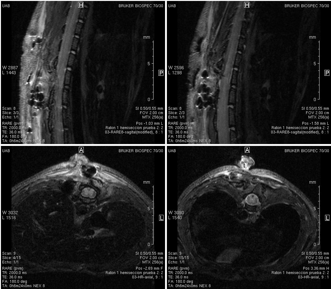
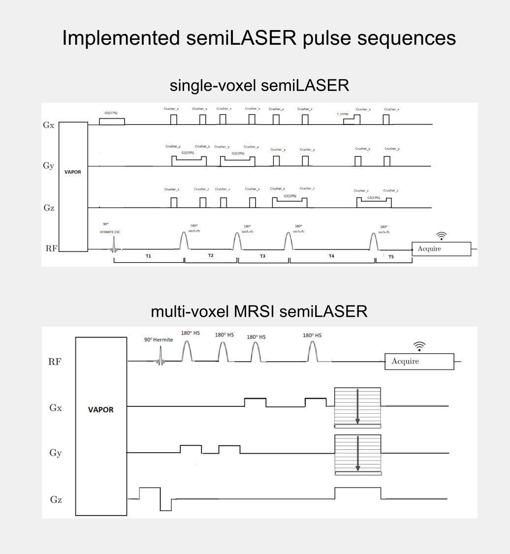
Although, the clinical utility of semiLASER has been acknowledged, the pre-clinical use and implementation of semiLASER still remains a less explored area. Our group has a long record of using MRSI in therapy response monitoring of a murine model glioblastoma (the GL261 cell line) using a commercially available MRSI acquisition sequence. In our efforts towards bridging the barriers between pre-clinical and clinical research, we have implemented the clinically verified semiLASER sequence on a pre-clinical 7T Bruker Biospec USR scanner running the ParaVision 5.1 software package, which provides a graphical user interface for sequence programming and data acquisition. The single and multi-voxel semiLASER sequences were implemented with the idea that the developments generated during this PhD project will be replicable by other interested users.
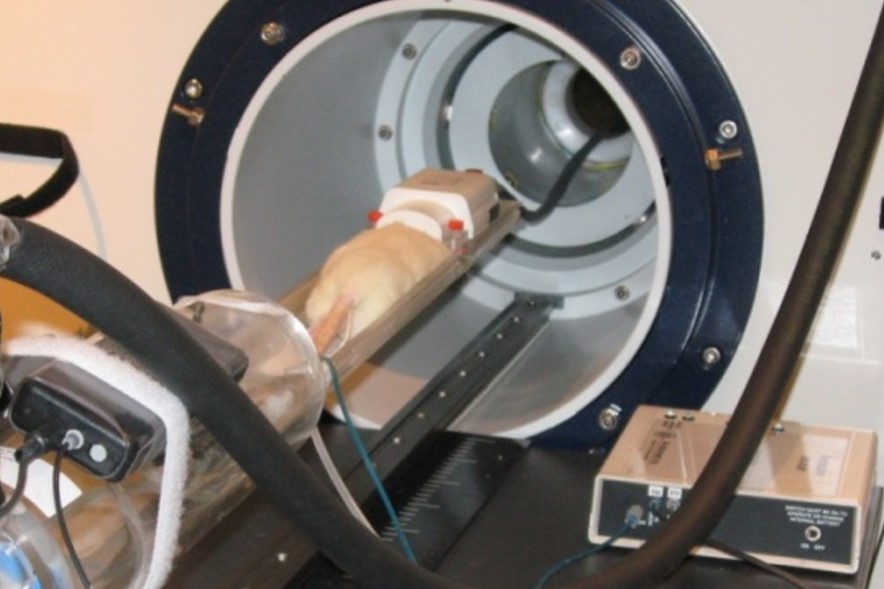
The implemented SV-semiLASER and MRSI-semiLASER sequences for preclinical acquisitions were optimised to perform high resolution MRSI of living mouse brain. For this, sequences were duly verified and tested first in phantoms and later in-vivo, in wild-type (wt) and tumor bearing (GL261) mice. To do so, the Bruker pulse sequence implementation was first studied in detail to become familiar with the Bruker programming environment and a test sequence PRESS_Slice to localize the slice dimension was developed by modifying the Bruker stock PRESS sequence for single voxel localization. After careful evaluation of test sequence results, the semiLASER single and multi-voxel sequences were also implemented using a similar strategy.
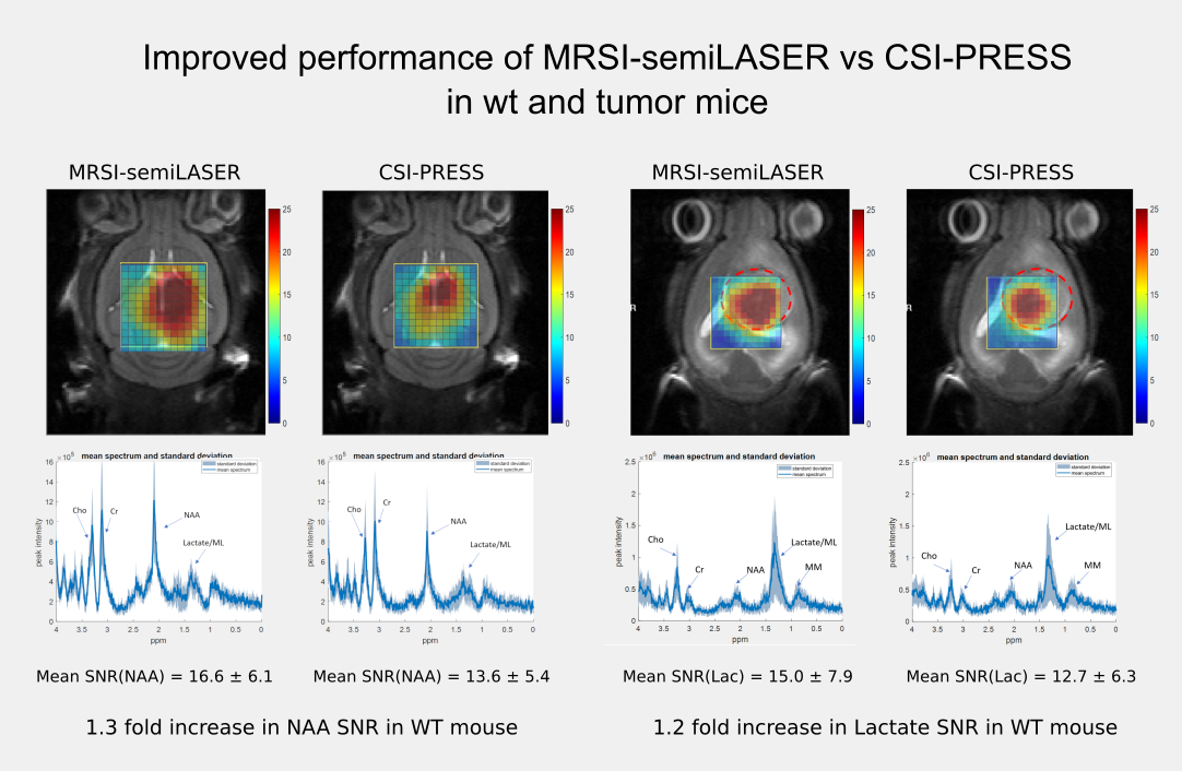
The implemented SV-semiLASER sequence provided a ca. 1.4-fold improvement in SNR in phantoms and ca. 1.3-fold improvement in SNR for in-vivo subjects, in comparison to the stock Bruker PRESS (single volume acquisition) sequence. The MRSI-semiLASER sequence resulted in a ca. 1.3-fold improvement of SNR in phantoms and in-vivo subjects compared to the stock Bruker CSI-PRESS sequence. Combined with phase encoding strategies and volume reduction methods, higher spatial resolution and SNR was achieved for the implemented MRSI-semiLASER. The quantification analysis of the results was done using MATLAB based post-processing tools specially designed to process Bruker datasets and solutions for a faster post processing pipeline were proposed. The single voxel MRSI-semiLASER sequences were further simulated using NMRSCOPE-B virtual simulator, a jMRUI plug-in which positively correlated with the experimental results. Preliminary nosological images obtained using MRSI-semiLASER datasets and the SpectraClassifier tool previously developed in our group, and trained with GL261 tumors using already available CSI-PRESS data, suggested those classifiers could be robust enough to recognize the tumor region acquired with the semi-LASER sequence. Still, classifiers may require retraining for the evaluation of response to therapy, which is an ongoing project within the group.
The thesis dissertation can be downloaded in PDF format using the link below or from the official TXD and Teseo repositories (currently in progress):
I would like to thank the financial support by the European Comission Marie Curie Initial Training Networks (ITN, call H2020-MSCA-ITN-2018, grant 813120 to project INSPiRE-MED); by the Ministry of Science and Insdustry (MCIN/AEI/10.13039/501100011033) (APC); and by Centro de Investigación Biomédica en Red—Bioingeniería, Biomateriales y Nanomedicina (CIBER-BBN http://www.ciber-bbn.es/en, CB06/01/0010), an initiative of the Instituto de Salud Carlos III (Spain) co-funded by the European Regional Development Fund (ERDF). I was recipient of a Marie Skłodowska-Curie early-stage researcher fellowship of the INSPiRE-MED project (Grant agreement ID: 813120)
Workshop limited to 4 participants (first come, first served)
Contact person:
Silvia Lope-Piedrafita, PhD ()
This course combines a comprehensive series of lectures on the technology of Magnetic resonance spectroscopy and imaging (MRS/MRI) with hands-on laboratory sessions to provide practical demonstrations of key concepts and procedures for preclinical studies.
Whether you are considering MRI as a research tool in your lab or just would like to learn more about MRI, this workshop addresses practical aspects of experimental MRI with laboratory animals and provide valuable hands-on experience on a 7 Tesla Bruker BioSpec spectrometer.
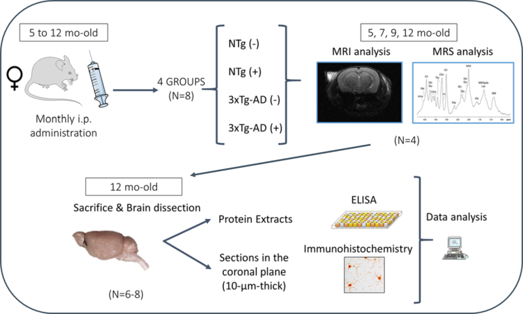
Progression of Alzheimer’s disease and effect of scFv-h3D6 immunotherapy in the 3xTg-AD mouse model: An in vivo longitudinal study using Magnetic Resonance Imaging and Spectroscopy by Güell-Bosch J, Lope-Piedrafita S, Esquerda-Canals G, Montoliu-Gaya L, and Villegas S. NMR in Biomedicine 33(5):e4263; DOI: 10.1002/nbm.4263.
Alzheimer’s disease (AD) is an incurable disease that affects most of the 47 million people estimated as living with dementia worldwide. The main histopathological hallmarks of AD are extracellular β-amyloid (Aβ) plaques and intracellular neurofibrillary tangles (NFTs) composed of hyperphosphorylated tau protein. In recent years, Aβ-immunotherapy has been revealed as a potential tool in AD treatment. One strategy consists of using single-chain variable fragments (scFvs), which avoids the fragment crystallizable (Fc) effects that are supposed to trigger a microglial response, leading to microhemorrhages and vasogenic edemas, as evidenced in clinical trials with bapineuzumab. The scFv-h3D6 generated by our research group derives from this monoclonal antibody, which targets the N-terminal of the Aβ peptide and recognizes monomers, oligomers and fibrils.
In this study, 3xTg-AD mice were intraperitoneally and monthly treated with 100 μg of scFv-h3D6 (a dose of ~3.3 mg/kg) or PBS, from 5 to 12 months of age (-mo), the age at which the mice were sacrificed and samples collected for histological and biochemical analyses. During treatments, four monitoring sessions using magnetic resonance imaging and spectroscopy (MRI/MRS) were performed at 5, 7, 9, and 12 months of age. MRI/MRS techniques allow, in a non-invasive manner, to draw an in vivo picture of concrete aspects of the pathology and to monitor its development across time. Compared with the genetic background, 3xTg-AD mice presented a smaller volume in almost all cerebral regions and ages examined, an increase in both the intra and extracellular Aβ1-42 at 12-mo, and an inflammation process at this age, in both the hippocampus (IL-6 and mIns) and cortex (IL-6). In addition, treatment with scFv-h3D6 partially recovered the values in brain volume, and Aβ, IL-6, and mIns concentrations, among others, encouraging further studies with this antibody fragment.
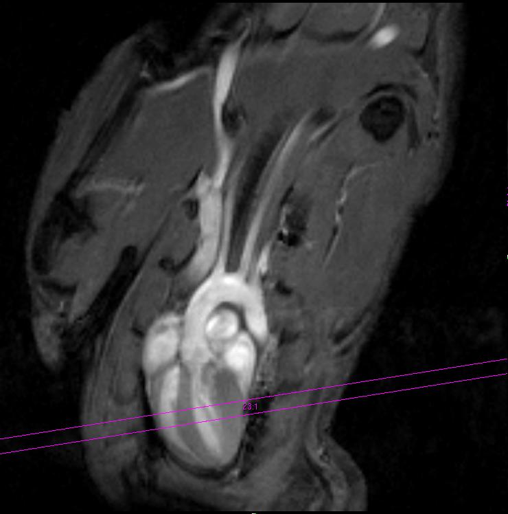
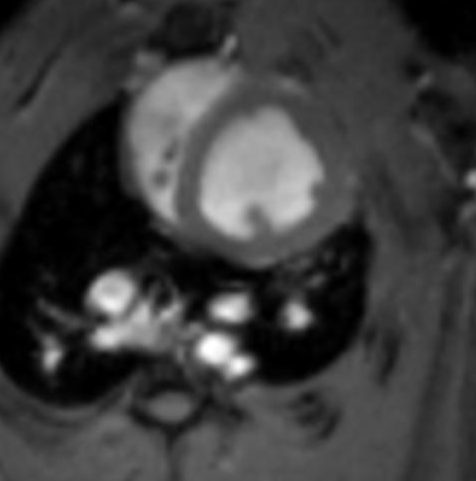
Simões, Rui V., Miquel E. Cabañas, Carla Loreiro, Miriam Illa, Fatima Crispi & Eduard Gratacós. 2018. Assessment of prenatal cerebral and cardiac metabolic changes in a rabbit model of fetal growth restriction based on 13C-labelled substrate infusions and ex vivo multinuclear HRMAS. PLOS ONE 13(12). e0208784. DOI: 10.1371/journal.pone.0208784
Background: We have used a previously reported rabbit model of fetal growth restriction (FGR), reproducing perinatal neurodevelopmental and cardiovascular impairments, to investigate the main relative changes in cerebral and cardiac metabolism of term FGR fetuses during nutrient infusion.
Methods: FGR was induced in 9 pregnant New Zealand rabbits at 25 days of gestation: one horn used as FGR, by partial ligation of uteroplacental vessels, and the contralateral as control (appropriate for gestation age, AGA). At 30 days of gestation, fasted mothers under anesthesia were infused i.v. with 1-13C-glucose (4 mothers), 2-13C-acetate (3 mothers), or not infused (2 mothers). Fetal brain and heart samples were quickly harvested and frozen down. Brain cortex and heart apex regions from 30 fetuses were studied ex vivo by HRMAS at 4°C, acquiring multinuclear 1D and 2D spectra. The data were processed, quantified by peak deconvolution or integration, and normalized to sample weight.
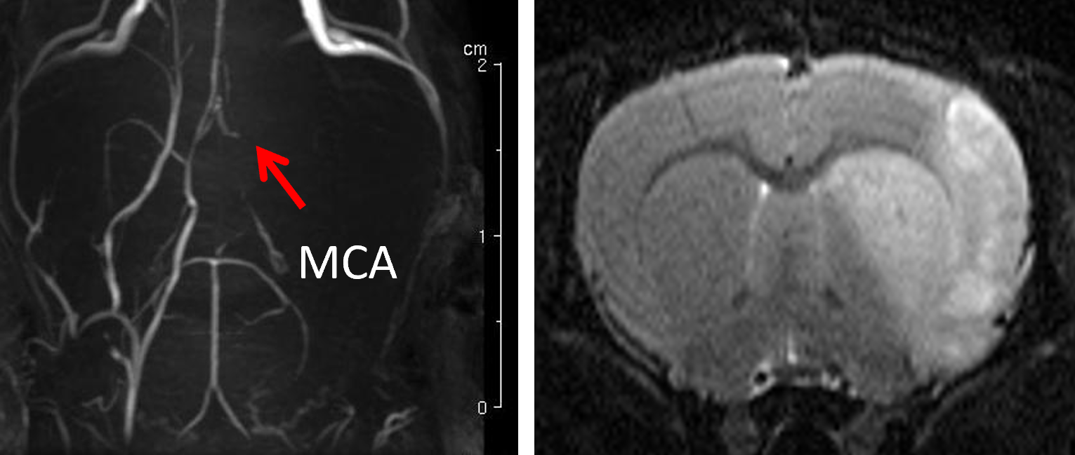
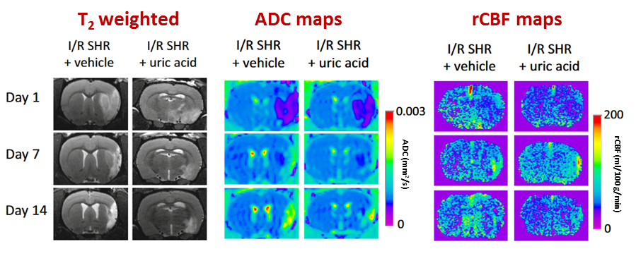
Jiménez-Xarrié, Elena, Belén Pérez, Ana Paula Dantas, Lídia Puertas-Umbert, Joan Martí-Fabregas, Ángel Chamorro, Anna Maria Planas, Elisabet Vila, Francesc Jiménez-Altayó. 2018. Uric Acid Treatment After Stroke Prevents Long-Term Middle Cerebral Artery Remodelling and Attenuates Brain Damage in Spontaneously Hypertensive Rats. Translational Stroke Research. DOI: 10.1007/s12975-018-0661-8
Hypertension is the most important modifiable risk factor for stroke and is associated with poorer post-stroke outcomes. The antioxidant uric acid is protective in experimental normotensive ischaemic stroke. However, it is unknown whether this treatment exerts long-term protection in hypertension. The authors aimed to evaluate the impact of transient intraluminal middle cerebral artery (MCA) occlusion (90min)/reperfusion (1–15 days) on brain and vascular damage progression in adult and spontaneously hypertensive rats (SHR) treated with uric acid. Ischaemic brain damage was assessed longitudinally with magnetic resonance imaging at the Nuclear Magnetic Resonance Service of the Universitat Autònoma de Barcelona.
In SHR rats, more severe brain damage and poorer neurofunctional outcomes were coupled to higher cortical cerebral blood flow at the onset of reperfusion, a transient increase in oxidative stress and long-lasting stroke-induced MCA hypertrophic remodelling. Thus, stroke promotes larger brain and vascular damage in hypertensive rats that persists for long-time. Uric acid administered during early reperfusion attenuated short- and long-term brain injuries in both normotensive and hypertensive rats, an effect that was associated with abolishment of the acute oxidative stress response and prevention of stroke-induced long lasting MCA remodelling in hypertension. These results suggest that uric acid might be an effective strategy to improve stroke outcomes in hypertensive subjects.
Montoliu-Gaya, Laia, Jofre Güell-Bosch, Gisela Esquerda-Canals, Alejandro R. Roda, Gabriel Serra-Mir, Silvia Lope-Piedrafita, José Luís Sánchez-Quesada & Sandra Villegas. 2018. Differential effects of apoE and apoJ mimetic peptides on the action of an anti-Aβ scFv in 3xTg-AD mice. Biochemical Pharmacology 155. 380–392. DOI: 10.1016/j.bcp.2018.07.012
Anti-Aβ immunotherapy has emerged as a promising approach to treat Alzheimer’s disease (AD). The single-chain variable fragment scFv-h3D6 is an anti-Aβ antibody fragment that lacks the Fc region, which is associated with the induction of microglial reactivity by the full-length monoclonal antibody bapineuzumab. ScFv-h3D6 was previously shown to restore the levels of apolipoprotein E (apoE) and apolipoprotein J (apoJ) in a tripletransgenic- AD (3xTg-AD) mouse model. Since apoE and apoJ play an important role in the development of AD, we aimed to study the in vivo effect of the combined therapy of scFv-h3D6 with apoE and apoJ mimetic peptides (MPs).
Workshop limited to 4 participants (first come, first served)
Contact person:
Silvia Lope-Piedrafita, PhD ()
This course combines a comprehensive series of lectures on the technology of Magnetic resonance spectroscopy and imaging (MRS/MRI) with hands-on laboratory sessions to provide practical demonstrations of key concepts and procedures for preclinical studies.
Whether you are considering MRI as a research tool in your lab or just would like to learn more about MRI, this workshop addresses practical aspects of experimental MRI with laboratory animals and provide valuable hands-on experience on a 7 Tesla Bruker BioSpec spectrometer.
García Martín, María Luisa & Pilar López Larrubia (eds.). 2018. Preclinical MRI. Methods in Molecular Biology series. Springer New York. DOI: 10.1007/978-1-4939-7531-0
This book was conceived with the idea of providing an update on a wide variety of preclinical MRI methods and protocols to help technicians and researchers interested in this technology. The basics of MRI physics are introduced, followed by chapters describing updated methodology and protocols for some standard and more advanced MRI techniques covering diffusion, perfusion, functional imaging, in-vivo spectroscopy (proton and heteronuclear), susceptibility contrast MRI… The book also contains some chapters where some applications of those methods are illustrated in animal models of several diseases including cancer, stroke and neurodegeneration. Protocols are described in a step-by-step approach, with interesting notes and tips at the end of each chapter, which -a priori- should allow the new worker to obtain successful results with the first attempt ;o) .