Tag Archives: mycolic acids

Precise characterization of mycobacterial cell wall lipid PTTM

Molecule confirmation and structure characterization of pentatriacontatrienyl mycolate in Mycobacterium smegmatisby M. Llorens-Fons, E. Julián, M. Luquin and M. Pérez-Trujillo. Chemistry and Physics of Lipids, 2018, Accepted Manuscript. DOI: https://doi.org/10.1016/j.chemphyslip.2017.12.006

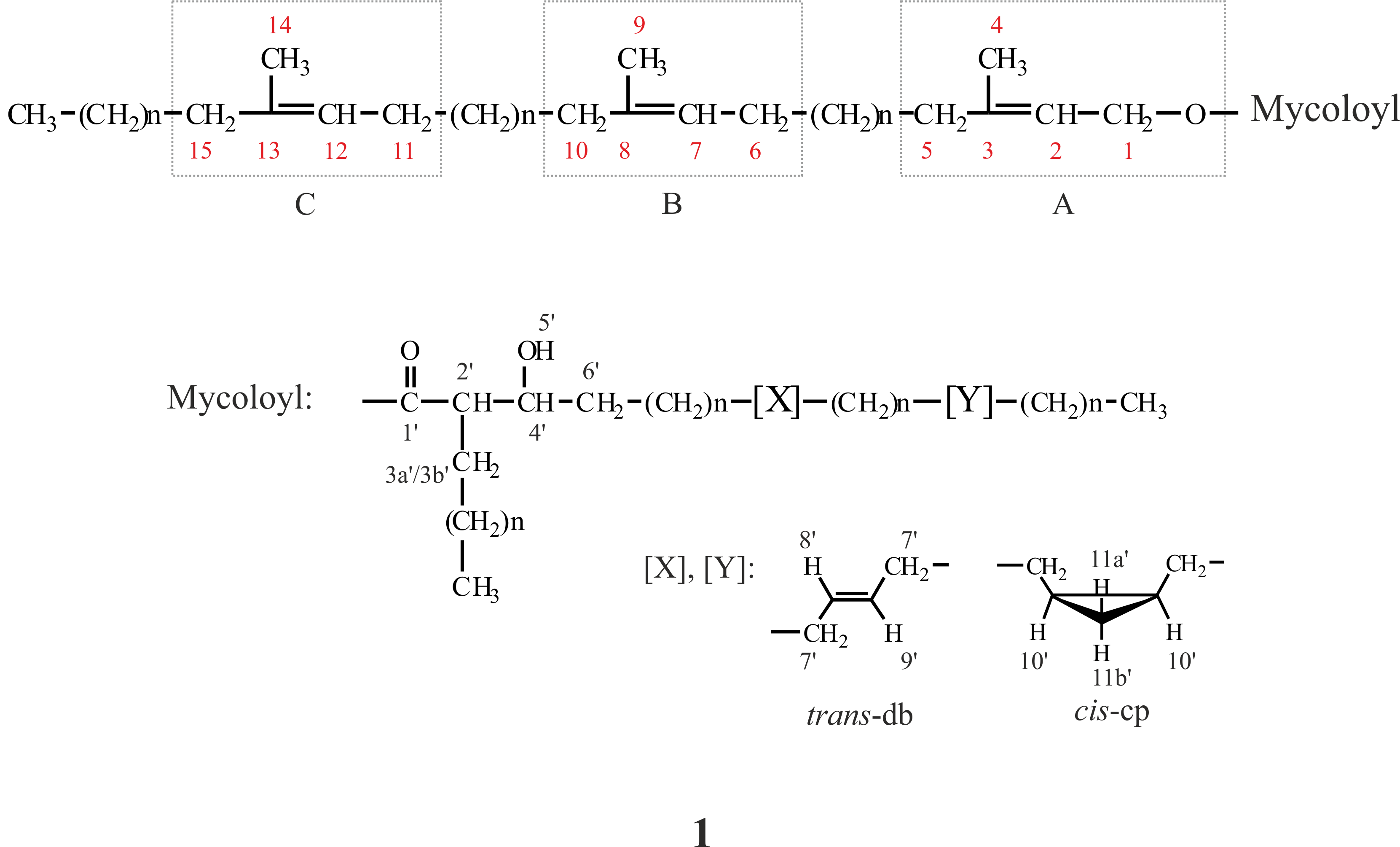
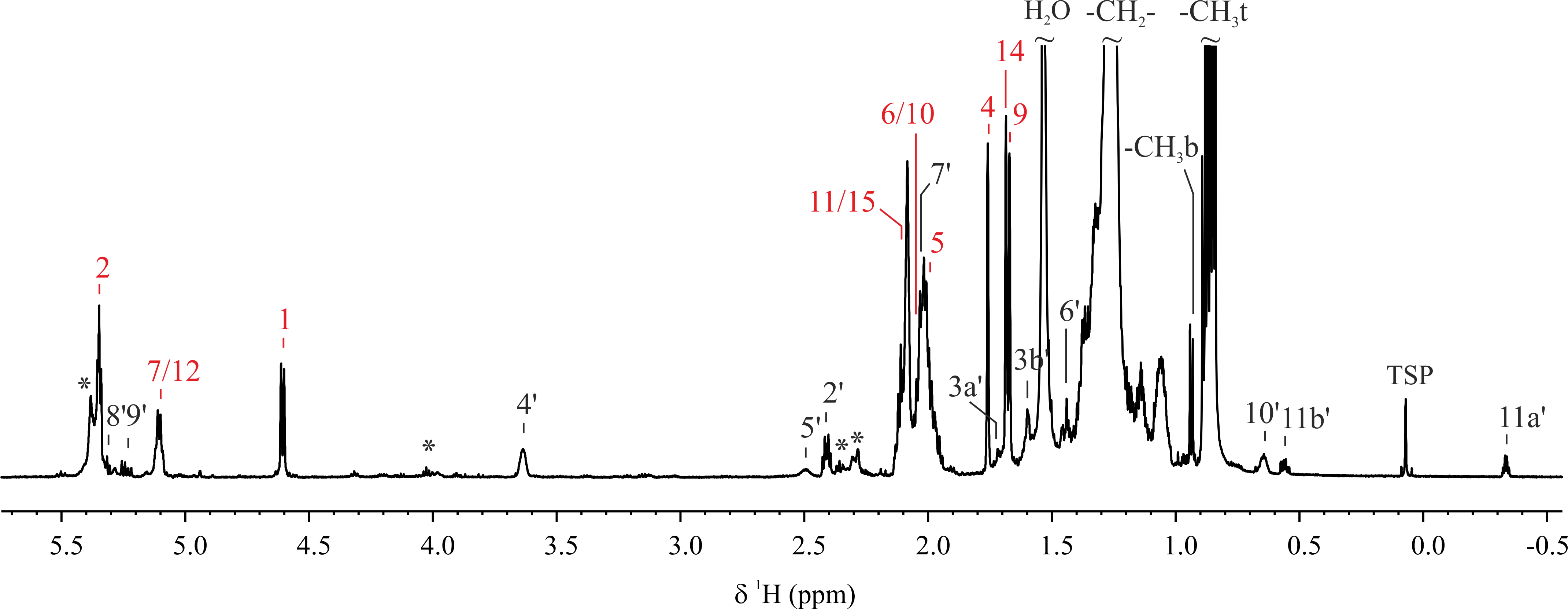
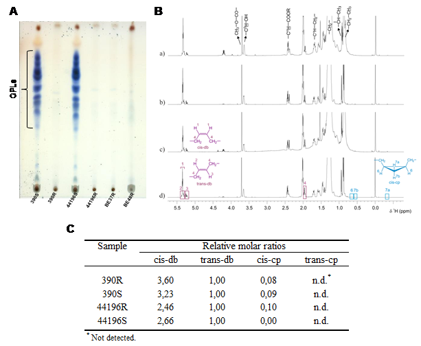
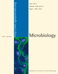
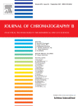
Mycobacterium smegmatis is often used to study the different components of mycobacterial cell wall. Mycolic acids are important components of mycobacterial cell wall that have been associated with virulence. Recently, a novel lipid containing mycolic acids has been described in M. smegmatis. However, some uncertainties regarding the structure of this molecule named mycolate ester wax have been reported. The objective of this work was to perform an in depth structural study of this molecule for its precise characterization. Using 1H and 13C NMR spectroscopy, the molecular structure of mycolate ester wax found in M. smegmatis has been elucidated. The characterization was complemented with MS analyses. This molecule is formed by a carbon chain with three methyl substituted olefinic units and a mycolate structure with trans double bonds and cis cyclopropane rings. The present molecular study will facilitate the detection and identification of pentatriacontatrienyl mycolate (PTTM) in future studies by the performance of a simple 1D 1H NMR experiment.

NMR identification of monstrous mycobacterial lipids in cell wall of Mycobacterium abcessus

” Trehalose polyphleates, external cell wall lipids in Mycobacterium abcessus, are associated with the formation of clumps with cording morphology, which have been associated with virulence” by M. Llorens-Fons, M. Pérez-Trujillo, E. Julián, C. Brambilla, F. Alcaide, T. F. Byrd and M. Luquin. Frontiers in Microbiology, 2017, 8:1402. DOI: https://dx.doi.org/10.3389/fmicb.2017.01402

Mycobacterium abscessus is a reemerging pathogen that causes pulmonary diseases similar to tuberculosis, which is caused by Mycobacterium tuberculosis. When grown in agar medium, M. abscessus strains generate rough (R) or smooth colonies (S). R morphotypes are more virulent than S morphotypes. In searching for the virulence factors responsible for this difference, R morphotypes have been found to form large aggregates (clumps) that, after being phagocytozed, result in macrophage death. Furthermore, the aggregates released to the extracellular space by damaged macrophages grow, forming unphagocytosable structures that resemble cords. In contrast, bacilli of the S morphotype, which do not form aggregates, do not damage macrophages after phagocytosis and do not form cords. Cording has also been related to the virulence of M. tuberculosis. A comparative study of the pattern and structure of mycolic acids was performed on R (cording) and S (non-cording) morphotypes derived from the same parent strains, and no differences were observed between morphotypes. Furthermore, cords formed by R morphotypes were disrupted with petroleum ether (PE), and the extracted lipids were analyzed by thin layer chromatography, nuclear magnetic resonance spectroscopy and mass spectrometry. Substantial amounts of trehalose polyphleates (TPP) were recovered as major lipids from PE extracts, and images obtained by transmission electron microscopy suggested that these lipids are localized to the external surfaces of cords and R bacilli. The structure of M. abscessus TPP was revealed to be similar to those previously described in Mycobacterium smegmatis. Although the exact role of TPP is unknown, our results demonstrated that TPP are not toxic by themselves and have a function in the formation of clumps and cords in M. abscessus, thus playing an important role in the pathogenesis of this species.


Mycobacteria clumping increase their capacity to damage macrophages

frontiersmicrobiology


“Mycobacteria clumping increase their capacity to damage macrophages” by C. Brambilla, M. Llorens-Fons, E. Julián, E. Noguera-Ortega, C. Tomàs-Martínez, M. Pérez-Trujillo, T. F. Byrd, F. Alcaide and M. Luquin.

Front. Microbiol. 7:1562.  DOI: 10.3389/fmicb.2016.01562

The rough morphotypes of non-tuberculous mycobacteria have been associated with the most severe illnesses in humans. This idea is consistent with the fact that Mycobacterium tuberculosis presents a stable rough morphotype. Unlike smooth morphotypes, the bacilli of rough morphotypes grow close together, leaving no spaces among them and forming large aggregates (clumps). Currently, the initial interaction of macrophages with clumps remains unclear. Thus, we infected J774 macrophages with bacterial suspensions of rough morphotypes of Mycobacterium abscessus containing clumps and suspensions of smooth morphotypes, primarily containing isolated bacilli. Using confocal laser scanning microscopy and electron microscopy, we observed clumps of at least 5 rough-morphotype bacilli inside the phagocytic vesicles of macrophages at 3 hours post-infection. These clumps grew within the phagocytic vesicles, killing 100% of the macrophages at 72 hours post-infection, whereas the proliferation of macrophages infected with smooth morphotypes remained unaltered at 96 hours post-infection. Thus, macrophages phagocytose large clumps, exceeding the bactericidal capacities of these cells. Furthermore, proinflammatory cytokines and granuloma-like structures were only produced by macrophages infected with rough morphotypes. Thus, the present study provides a foundation for further studies that consider mycobacterial clumps as virulence factors.

fig8

Figure. Content of GPL and structure of mycolic acids. (A) 1-D TLC analysis of the crude lipid extracts of M. abscessus strains. (B) 1H-NMR spectra of purified mycolic acid methyl esters from M. abscessus. (C) Relative molar ratios of molecular moieties cis-db, trans-db, cis-cp and trans-cp of mycolic acid methyl esters from M. abscessus.

Tuberculosis mycobacterium and microscopic cords formation. Related to their virulence

“Cyclopropanation of α-mycolic acids is not required for cording in non-tuberculous mycobacteria” Cecilia Brambilla, Alejandro Sánchez-Chardi, Míriam Pérez-Trujillo, Esther Julián, Marina Luquin.  Microbiology, March 2012. DOI: 10.1099/mic.0.057919-0

Mycobacterium tuberculosis and Mycobacterium bovis are the principal agents responsible for human tuberculosis and tuberculosis in domestic and wild animals. Both form microscopic cords, a phenotypic characteristic that has been related to their virulence. The compounds responsible for cording are unknown, but a recent work has shown that cording could be related to the fine structure of α-mycolic acids. Continue reading Tuberculosis mycobacterium and microscopic cords formation. Related to their virulence

Revisited mycolic acid pattern of Mycobacterium confluentis using thin-layer chromatography

“Revisited mycolic acid pattern of Mycobacterium confluentis using thin-layer chromatography”, by Silvia Secanella-Fandos, Marina Luquin, Míriam Pérez-Trujillo, Esther Julián; Journal of Chromatography B, Article in Press (2011). doi:10.1016/j.jchromb.2011.08.001

The profile of mycolic acids from Mycobacterium confluentis has not been adequately published. However, the definition of the composition of mycolic acids is a critical element for describing new mycobacterial species. Thus, an erroneously published profile can lead to confusing citations. Continue reading Revisited mycolic acid pattern of Mycobacterium confluentis using thin-layer chromatography