 “Dilated cardiomyopathy and mitochondrial dysfunction in Sirt1-deficient mice: A role for Sirt1-Mef2 in adult heart”
by A. Planavila, E. Dominguez, M. Navarro, M. Vinciguerra, R. Iglesias, M. Giralt, S. Lope-Piedrafita, J. Ruberte, F. Villarroya. Journal of Molecular and Cellular Cardiology 53 (2012) 521-531. DOI: 10.1016/j.yjmcc.2012.07.019
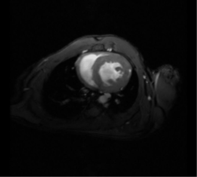 The protein deacetylase Sirtuin-1 (Sirt1) is involved in the cardiac hypertrophic responses and cardiac embryo morphogenesis. However, the physiological function of Sirt1 deficiency in the postnatal development of the heart remains to be characterized. The aim of this study was to investigate the relevance of Sirt1 in the development and function of the myocardium by using complementary techniques, such as gene expression, immunoblotting, immunohistochemistry, histological and electron microscopy examinations, and in vivo cardiac magnetic resonance imaging (MRI).
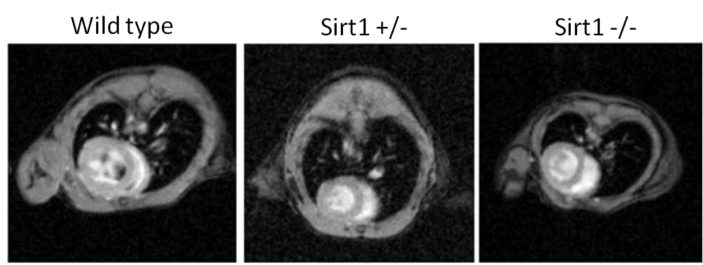
MRI data indicated that Sirt1 deficiency led to an enlargement of the left ventricular (LV) chamber. The ratio between left ventricular wall and septal thickness versus left ventricular chamber radius, a measure of dilatation referred as the H/R ratio, was decreased in Sirt1+/− (0.20 ± 0.02) and Sirt1−/− mice (0.21 ± 0.03) compared to wild-type mice (0.42 ± 0.07).