Workshop limited to 4 participants (first come, first served)
Contact person:
Silvia Lope-Piedrafita, PhD ()
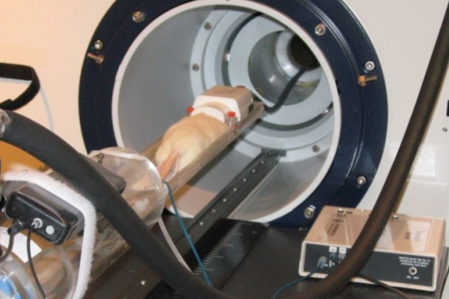
This course combines a comprehensive series of lectures on the technology of Magnetic resonance spectroscopy and imaging (MRS/MRI) with hands-on laboratory sessions to provide practical demonstrations of key concepts and procedures for preclinical studies.
Whether you are considering MRI as a research tool in your lab or just would like to learn more about MRI, this workshop addresses practical aspects of experimental MRI with laboratory animals and provide valuable hands-on experience on a 7 Tesla Bruker BioSpec spectrometer.
The Specialised Group of NMR of the Spanish Royal Society of Chemistry (GERMN, RSEQ) organizes the XV Manuel Rico NMR Summer School in Jaca from 19th-23th June 2023.
This well-established bi-annual summer course is aimed at PhD students, postdocs, technical staff of NMR facilities and, in general, to researchers from academy and industry interested in deepening their understanding of NMR. The course covers theoretical aspects, state-of-the-art methods and applications in fields as diverse as Molecular Chemistry, Materials, Biology, Medicine, and Pharmaceutical Industry, including solution-state, and solid-state NMR techniques, as well as MRI techniques.
Silvia Lope, SeRMN staff, will be teaching a class in “Magnetic Resonance Imaging”.
The Specialised Group of NMR of the Spanish Royal Society of Chemistry (GERMN, RSEQ) organizes the XIV Manuel Rico NMR Summer School in Jaca from 19th-24th June 2022.
This well-established bi-annual summer course is aimed at PhD students, postdocs, technical staff of NMR facilities and, in general, to researchers from academy and industry interested in deepening their understanding of NMR. The course covers theoretical aspects, state-of-the-art methods and applications in fields as diverse as Molecular Chemistry, Materials, Biology, Medicine, and Pharmaceutical Industry, including solution-state, and solid-state NMR techniques, as well as MRI techniques.
Silvia Lope, SeRMN staff, will be teaching a class in “Magnetic Resonance Imaging”.
End of pre-registration April 12th, 2022
Start of Registration: April 30th, 2022
End of Registration: May 17th, 2022
For detailed information please visit their webpage https://rmnjaca22.iqfr.csic.es/
Workshop limited to 4 participants (first come, first served)
Contact person:
Silvia Lope-Piedrafita, PhD ()
This course combines a comprehensive series of lectures on the technology of Magnetic resonance spectroscopy and imaging (MRS/MRI) with hands-on laboratory sessions to provide practical demonstrations of key concepts and procedures for preclinical studies.
Whether you are considering MRI as a research tool in your lab or just would like to learn more about MRI, this workshop addresses practical aspects of experimental MRI with laboratory animals and provide valuable hands-on experience on a 7 Tesla Bruker BioSpec spectrometer.
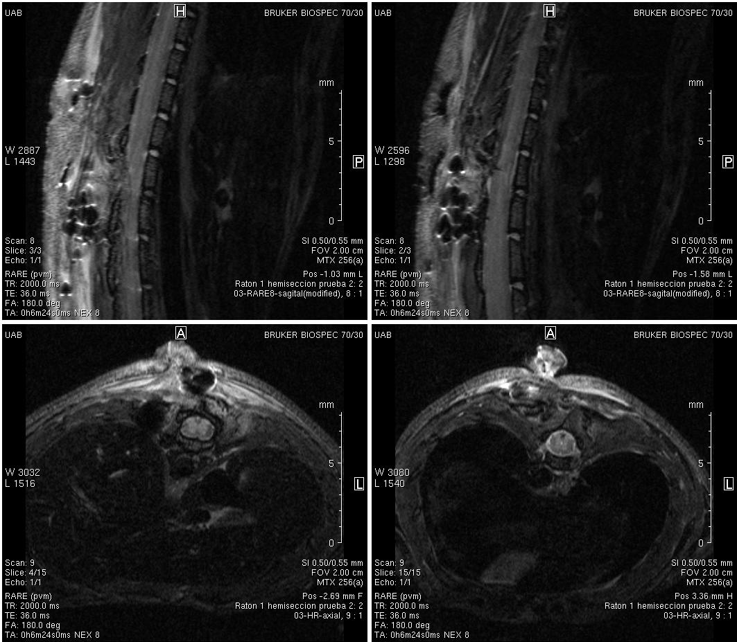
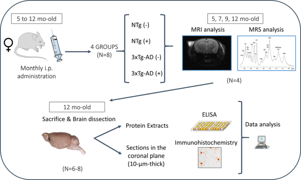
Progression of Alzheimer’s disease and effect of scFv-h3D6 immunotherapy in the 3xTg-AD mouse model: An in vivo longitudinal study using Magnetic Resonance Imaging and Spectroscopy by Güell-Bosch J, Lope-Piedrafita S, Esquerda-Canals G, Montoliu-Gaya L, and Villegas S. NMR in Biomedicine 33(5):e4263; DOI: 10.1002/nbm.4263.
Alzheimer’s disease (AD) is an incurable disease that affects most of the 47 million people estimated as living with dementia worldwide. The main histopathological hallmarks of AD are extracellular β-amyloid (Aβ) plaques and intracellular neurofibrillary tangles (NFTs) composed of hyperphosphorylated tau protein. In recent years, Aβ-immunotherapy has been revealed as a potential tool in AD treatment. One strategy consists of using single-chain variable fragments (scFvs), which avoids the fragment crystallizable (Fc) effects that are supposed to trigger a microglial response, leading to microhemorrhages and vasogenic edemas, as evidenced in clinical trials with bapineuzumab. The scFv-h3D6 generated by our research group derives from this monoclonal antibody, which targets the N-terminal of the Aβ peptide and recognizes monomers, oligomers and fibrils.
In this study, 3xTg-AD mice were intraperitoneally and monthly treated with 100 μg of scFv-h3D6 (a dose of ~3.3 mg/kg) or PBS, from 5 to 12 months of age (-mo), the age at which the mice were sacrificed and samples collected for histological and biochemical analyses. During treatments, four monitoring sessions using magnetic resonance imaging and spectroscopy (MRI/MRS) were performed at 5, 7, 9, and 12 months of age. MRI/MRS techniques allow, in a non-invasive manner, to draw an in vivo picture of concrete aspects of the pathology and to monitor its development across time. Compared with the genetic background, 3xTg-AD mice presented a smaller volume in almost all cerebral regions and ages examined, an increase in both the intra and extracellular Aβ1-42 at 12-mo, and an inflammation process at this age, in both the hippocampus (IL-6 and mIns) and cortex (IL-6). In addition, treatment with scFv-h3D6 partially recovered the values in brain volume, and Aβ, IL-6, and mIns concentrations, among others, encouraging further studies with this antibody fragment.
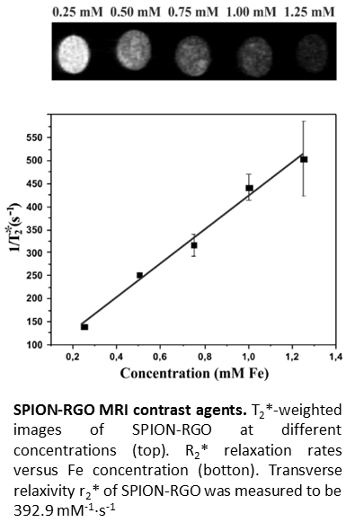
Magnetic resonance imaging (MRI) is a useful tool for disease diagnosis
and treatment monitoring. Superparamagnetic iron oxide nanoparticles (SPION)
show good performance as transverse relaxation (T2) contrast agents, thus
facilitating the interpretation of the acquired images. Attachment of SPION
onto nanocarriers prevents their agglomeration, improving the circulation time
and efficiency. Graphene derivatives, such as graphene oxide (GO) and reduced
graphene oxide (RGO), are appealing nanocarriers since they have both high
surface area and functional moieties that make them ideal substrates for the
attachment of nanoparticles. A fast, simple, and environmentally friendly
microwave-assisted approach for the synthesis of SPION-RGO hybrids has been demonstrated
in this study. Different iron precursor/GO ratios were used leading to SPION,
with a median diameter of 7.1 nm, homogeneously distributed along the RGO
surface. Good relaxivity (r2*) values were obtained in MRI studies and no
significant toxicity was detected within in vitro tests following GL261 glioma
and J774 macrophage-like cells for 24 h with SPION-RGO, demonstrating the
applicability of the hybrids as T2-weighted MRI contrast agents.
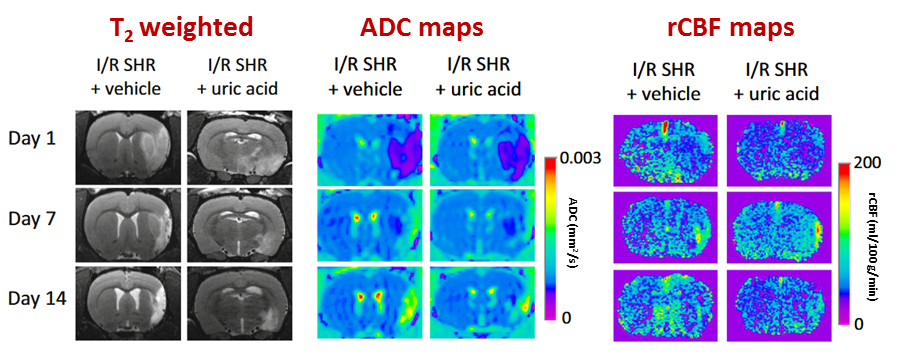
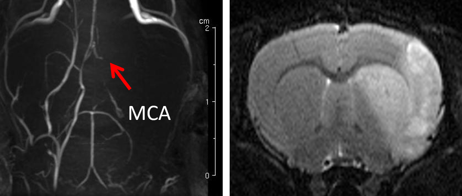
Jiménez-Xarrié, Elena, Belén Pérez, Ana Paula Dantas, Lídia Puertas-Umbert, Joan Martí-Fabregas, Ángel Chamorro, Anna Maria Planas, Elisabet Vila, Francesc Jiménez-Altayó. 2018. Uric Acid Treatment After Stroke Prevents Long-Term Middle Cerebral Artery Remodelling and Attenuates Brain Damage in Spontaneously Hypertensive Rats. Translational Stroke Research. DOI: 10.1007/s12975-018-0661-8
Hypertension is the most important modifiable risk factor for stroke and is associated with poorer post-stroke outcomes. The antioxidant uric acid is protective in experimental normotensive ischaemic stroke. However, it is unknown whether this treatment exerts long-term protection in hypertension. The authors aimed to evaluate the impact of transient intraluminal middle cerebral artery (MCA) occlusion (90min)/reperfusion (1–15 days) on brain and vascular damage progression in adult and spontaneously hypertensive rats (SHR) treated with uric acid. Ischaemic brain damage was assessed longitudinally with magnetic resonance imaging at the Nuclear Magnetic Resonance Service of the Universitat Autònoma de Barcelona.
In SHR rats, more severe brain damage and poorer neurofunctional outcomes were coupled to higher cortical cerebral blood flow at the onset of reperfusion, a transient increase in oxidative stress and long-lasting stroke-induced MCA hypertrophic remodelling. Thus, stroke promotes larger brain and vascular damage in hypertensive rats that persists for long-time. Uric acid administered during early reperfusion attenuated short- and long-term brain injuries in both normotensive and hypertensive rats, an effect that was associated with abolishment of the acute oxidative stress response and prevention of stroke-induced long lasting MCA remodelling in hypertension. These results suggest that uric acid might be an effective strategy to improve stroke outcomes in hypertensive subjects.
Montoliu-Gaya, Laia, Jofre Güell-Bosch, Gisela Esquerda-Canals, Alejandro R. Roda, Gabriel Serra-Mir, Silvia Lope-Piedrafita, José Luís Sánchez-Quesada & Sandra Villegas. 2018. Differential effects of apoE and apoJ mimetic peptides on the action of an anti-Aβ scFv in 3xTg-AD mice. Biochemical Pharmacology 155. 380–392. DOI: 10.1016/j.bcp.2018.07.012
Anti-Aβ immunotherapy has emerged as a promising approach to treat Alzheimer’s disease (AD). The single-chain variable fragment scFv-h3D6 is an anti-Aβ antibody fragment that lacks the Fc region, which is associated with the induction of microglial reactivity by the full-length monoclonal antibody bapineuzumab. ScFv-h3D6 was previously shown to restore the levels of apolipoprotein E (apoE) and apolipoprotein J (apoJ) in a tripletransgenic- AD (3xTg-AD) mouse model. Since apoE and apoJ play an important role in the development of AD, we aimed to study the in vivo effect of the combined therapy of scFv-h3D6 with apoE and apoJ mimetic peptides (MPs).
Workshop limited to 4 participants (first come, first served)
Contact person:
Silvia Lope-Piedrafita, PhD ()
This course combines a comprehensive series of lectures on the technology of Magnetic resonance spectroscopy and imaging (MRS/MRI) with hands-on laboratory sessions to provide practical demonstrations of key concepts and procedures for preclinical studies.
Whether you are considering MRI as a research tool in your lab or just would like to learn more about MRI, this workshop addresses practical aspects of experimental MRI with laboratory animals and provide valuable hands-on experience on a 7 Tesla Bruker BioSpec spectrometer.
 by Llenas M, Sandoval S, Costa PM, Oró-Solé J,