Tag Archives: HRMAS

In vivo MRS and ex vivo HRMAS in an Ischemic Rat Stroke Model

JCBMcover
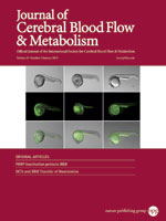
“In vivo and ex vivo Magnetic Resonance Spectroscopy of the Infarct and the Subventricular Zone in Experimental Stroke” by E. Jiménez-Xarrié, M. Davila, S. Gil-Perotín, A. Jurado-Rodríguez, A.P. Candiota, R. Delgado-Mederos, S. Lope-Piedrafita, J.M. García-Verdugo, C. Arús, J. Martí-Fàbregas. Journal of Cerebral Blood Flow & Metabolism, 2015, 35:828–834. DOI: 10.1038/jcbfm.2014.257

Ischemic stroke changes the metabolic pattern in the infarct area and also in other regions such as the ipsilateral subventricular zone (SVZi) where neural progenitor cells (NPCs) proliferation is enhanced in the mammalian and human brains. Magnetic resonance spectroscopy (MRS) provides metabolic information in vivo. With regard to NPCs proliferation, a resonance at 1.28 ppm has been described as an in vivo MRS biomarker of NPCs in the hippocampus of rats and humans. Continue reading In vivo MRS and ex vivo HRMAS in an Ischemic Rat Stroke Model

Free sugars quantification in intact plant tissue by HR-MAS NMR.

Planta - Journal cover“Assessment of a 1H high-resolution magic angle spinning NMR spectroscopy procedure for free sugars quantification in intact plant tissue” Teresa Delgado-Goñi, Sonia Campo, Juana Martín-Sitjar, Miquel E. Cabañas, Blanca San Segundo, Carles Arús; Planta, 238:397-413, 2013 (PDF file). DOI: 10.1007/s00425-013-1924-y

In most plants, sucrose is the primary product of photosynthesis, the transport form of assimilated carbon, and also one of the main factors determining sweetness in fresh fruits. Traditional methods for sugar quantification (mainly sucrose, glucose and fructose) require obtaining crude plant extracts, which sometimes involve substantial sample manipulation, making the process time-consuming and increasing the risk of sample degradation. Here, we describe and validate a fast method to determine sugar content in intact plant tissue by using high-resolution magic angle spinning nuclear magnetic resonance spectroscopy (HR-MAS NMR). Continue reading Free sugars quantification in intact plant tissue by HR-MAS NMR.

How does HR-MAS spin rate affect the spectral pattern of mobile lipids?

“Influence of the spinning rate in the HR-MAS pattern of mobile lipids in C6 glioma cells and in artificial oil bodies” Juana Martín-Sitjar, Teresa Delgado-Goñi, Miquel E. Cabañas, Jason Tzen, Carles Arús; Magnetic Resonance Materials in Physics, Biology and Medicine, 2012, 25(6):487-496. DOI: 10.1007/s10334-012-0327-6

Object: To evaluate how spinning rate affects mobile lipid (ML) resonances visibility in HR-MAS spectra of C6 glioma cells and artificial oil bodies (AOB), as models of cytosolic lipid droplets. Materials and methods: Using C6 cells and AOB of two different sizes, 780 ± 580 and 240 ± 293 nm, as models, we acquired HR-MAS pulse and acquire spectra at different spinning rates between 500 and 15,000 Hz, all at 37 °C. Continue reading How does HR-MAS spin rate affect the spectral pattern of mobile lipids?

HR-MAS: a key technique to study Nanoparticles

“Phosphine Stabilized Ruthenium Nanoparticles: the Effect of the Nature of the Ligand in Catalysis” David González-Gálvez, Pau Nolis, Karine Philippot, Bruno Chaudret and Piet W. N. M. van Leeuwen. ACS Catalysis, Volume 2, Pages 317-321, January 2012 DOI: 10.1021/cs200633k

Various ligands not forming monometallic complexes were used for Ru nanoparticle stabilization, enabling the control of size, shape and electronic properties. HRMAS NMR spectroscopy allowed us to study surface-bound molecules, evidencing ligand hydrogenation and decomposition of THF during the RuNP synthesis. Continue reading HR-MAS: a key technique to study Nanoparticles