“Dimeth yl sulfoxide (DMSO) as a potential contrast agent for brain tumors”, by Delgado-Goñi T, Martín-Sitjar J, Simões RV, Acosta M, Lope-Piedrafita S, Arús C.; NMR in Biomedicine. 2012.DOI: 10.1002/nbm.2832.
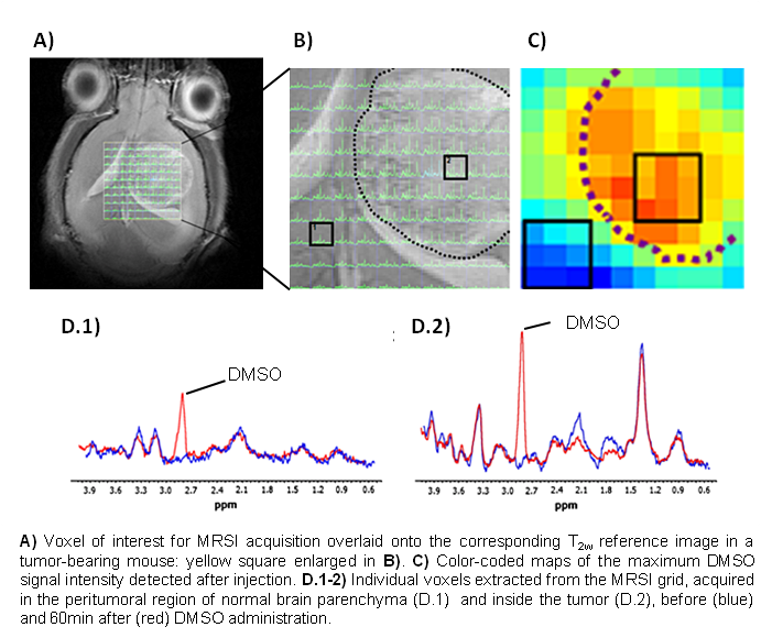
Identifying the type of brain tumor helps doctors determine the most appropriate course of treatment. Brain tumor diagnosis usually involves a neurological examination, brain scans, and/or an analysis of the brain tissue. Nowadays, the biopsy, although it is a very invasive procedure, is the most accurate method of obtaining a diagnosis. It would be very important to find alternative non-invasive methods which would also provide a reliable diagnosis, and magnetic resonance spectroscopy (MRS) and magnetic resonance spectroscopic imaging (MRSI) may have a significant role to play here as they provide the metabolic profile of these abnormal masses in a non-invasive manner. Moreover, the used of contrast agents may further improve tumor classification. Within this framework, in this study we evaluated DMSO as a potential contrast agent for detecting brain tumors. DMSO was administrated to normal and tumor-bearing mice and they were investigated by MRS and MRSI. Differential DMSO accumulation and wash-out kinetics were observed in normal versus tumor-afflicted brain parenchyma. MRSI maps of time-course DMSO changes revealed clear hot-spots of differential spatial accumulation even in tumors that did not enhance in T1w-images post gadolinium injection. Our results indicate a potential role for DMSO as a contrast agent for brain tumor detection, and possibly for monitoring their heterogeneities, for example due to progression or to therapy response.