 “In vivo and ex vivo Magnetic Resonance Spectroscopy of the Infarct and the Subventricular Zone in Experimental Stroke” by E. Jiménez-Xarrié, M. Davila, S. Gil-Perotín, A. Jurado-Rodríguez, A.P. Candiota, R. Delgado-Mederos, S. Lope-Piedrafita, J.M. García-Verdugo, C. Arús, J. Martí-Fàbregas. Journal of Cerebral Blood Flow & Metabolism, 2015, 35:828–834. DOI: 10.1038/jcbfm.2014.257
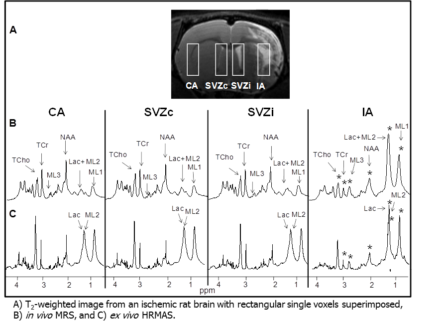
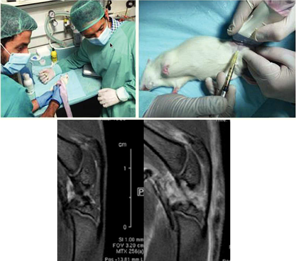
Ischemic stroke changes the metabolic pattern in the infarct area and also in other regions such as the ipsilateral subventricular zone (SVZi) where neural progenitor cells (NPCs) proliferation is enhanced in the mammalian and human brains. Magnetic resonance spectroscopy (MRS) provides metabolic information in vivo. With regard to NPCs proliferation, a resonance at 1.28 ppm has been described as an in vivo MRS biomarker of NPCs in the hippocampus of rats and humans. However, many authors disagree with this interpretation as this resonance has been found in different cell types and during different developmental stages corresponding to mobile lipids (MLs). High-resolution magic-angle spinning (HRMAS) also provides metabolic information, and moreover, with higher sensitivity and spectral resolution than MRS, however, it is used ex vivo and postmortem metabolic changes could affect the final metabolic pattern. Therefore, in this study, in vivo MRS and ex vivo HRMAS were used as complementary techniques to better characterize the metabolic pattern of the infarct and the NPCs in the ipsilateral subventricular zone.