All posts by Miriam

31P-NMR Metabolomics Revealed Species-Specific Use of Phosphorous in Trees

31P-NMR Metabolomics Revealed Species-Specific Use of Phosphorous in Trees of a French Guiana Rainforest, by Gargallo-Garriga, A.; Sardans, J.; Llusià, J.; Peguero, G.; Asensio, D.; Ogaya, R.; Urbina, I.; Langenhove, L.V.; Verryckt, L.T.; Courtois, E.A.; Stahl, C.; Grau, O.; Urban, O.; Janssens, I.A.; Nolis, P.; Pérez-Trujillo, M.; Parella, T.; Peñuelas, J.  Molecules 202025, 3960. https://doi.org/10.3390/molecules25173960

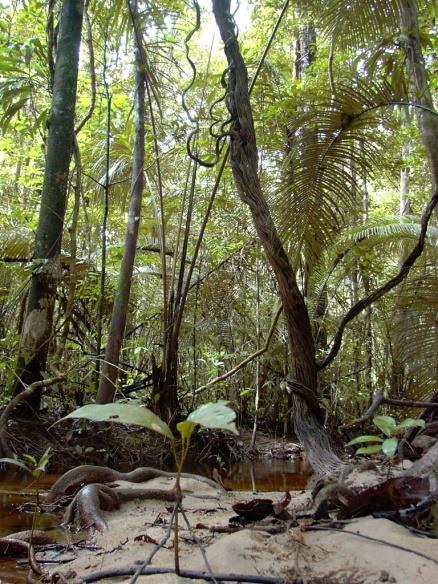
Productivity of tropical lowland moist forests is often limited by availability and functional allocation of phosphorus (P) that drives competition among tree species and becomes a key factor in determining forestall community diversity. We used non-target 31P-NMR metabolic profiling to study the foliar P-metabolism of trees of a French Guiana rainforest. The objective was to test the hypotheses that P-use is species-specific, and that species diversity relates to species P-use and concentrations of P-containing compounds, including inorganic phosphates, orthophosphate monoesters and diesters, phosphonates and organic polyphosphates. We found that tree species explained the 59% of variance in 31P-NMR metabolite profiling of leaves. A principal component analysis showed that tree species were separated along PC 1 and PC 2 of detected P-containing compounds, which represented a continuum going from high concentrations of metabolites related to non-active P and P-storage, low total P concentrations and high N:P ratios, to high concentrations of P-containing metabolites related to energy and anabolic metabolism, high total P concentrations and low N:P ratios. These results highlight the species-specific use of P and the existence of species-specific P-use niches that are driven by the distinct species-specific position in a continuum in the P-allocation from P-storage compounds to P-containing molecules related to energy and anabolic metabolism.

This article belongs to the Special Issue:

https://www.mdpi.com/journal/molecules/special_issues/nmr_metabolomics

Recyclable Mesoporous Organosilica Nanoparticles (MSN) for Asymmetric Organocatalysis

Li, Hao, Míriam Pérez-Trujillo, Xavier Cattoën & Roser Pleixats. 2019. Recyclable Mesoporous Organosilica Nanoparticles Derived from Proline-Valinol Amides for Asymmetric Organocatalysis. ACS Sustainable Chemistry & Engineering 7(17). 14815-14828. DOI: 10.1021/acssuschemeng.9b02838

This is the first report on the obtention of functionalized MSN by a co-condensation procedure with a structurally complex chiral precursor. The functionalized MSN have been characterized by elemental analysis, 29Si and 13C CP MAS NMR, transmission electron microscopy, scanning electron microscopy, N2-sorption measurements, dynamic light scattering, ζ-potential, and powder X-ray diffraction. We have evaluated the activity of these materials as recyclable catalysts in the asymmetric aldol reaction. The use of organosilica nanoparticles reduces the problems of diffusion and low reaction rates encountered with bulk organosilicas.

Exopolysaccharides from olive brines could reduce the adhesion of ETEC K88 to intestinal epithelial cells

Zhu, Yanan, Gemma González-Ortiz, Rufino Jiménez-Díaz, Míriam Pérez-Trujillo, Teodor Parella, Paola López-Colom & Susana María Martín-Orúe. 2018. Exopolysaccharides from olive brines could reduce the adhesion of ETEC K88 to intestinal epithelial cells. Food & Function 9(7). 3884–3894. DOI: https://doi.org/10.1039/c8fo00690c

This study aims to explore the biological functions of the isolated exopolysaccharides (EPSs) produced during the industrial fermentation of olives against enterotoxigenic E. coli (ETEC) K88. Exopolysaccharides were isolated from five industrial fermenters. Analysis of their monosaccharide composition by GLC revealed that the main components were glucose (27%–50%) and galactose (23%–33%) followed by rhamnose (4–23%) and arabinose (6–17%). The 1H NMR spectrum showed a very similar profile between samples, and a more in-depth analysis revealed the presence of an α-pyranose in the form of α-D-Glcp-(1→) and two different α-furanoses, with chemicals shift values, suggesting the presence of α-D-Glcf and α-D-Galf. Miniaturized in vitro tests demonstrated the ability of EPS samples to attach specifically to ETEC K88 (P < 0.05) with variable intensities. The competition test did not show the ability to block the ETEC K88 adhesion to IPEC-J2 cells; however, in the displacement test, all EPS samples were shown to effectively remove the pathogens attached to the cells (P < 0.01).

These results suggest that the EPSs produced during the fermentation of table green olives could interfere with the attachment of opportunistic pathogens onto the intestinal epithelial cells. This would open the possibility of novel functional properties for this traditional Mediterranean fermented food and for the isolated EPSs as candidates for nutraceutics to be used in human and/or animal diets in the prevention and treatment of ETEC diarrhoea.

Precise characterization of mycobacterial cell wall lipid PTTM

Molecule confirmation and structure characterization of pentatriacontatrienyl mycolate in Mycobacterium smegmatisby M. Llorens-Fons, E. Julián, M. Luquin and M. Pérez-Trujillo. Chemistry and Physics of Lipids, 2018, Accepted Manuscript. DOI: https://doi.org/10.1016/j.chemphyslip.2017.12.006

Mycobacterium smegmatis is often used to study the different components of mycobacterial cell wall. Mycolic acids are important components of mycobacterial cell wall that have been associated with virulence. Recently, a novel lipid containing mycolic acids has been described in M. smegmatis. However, some uncertainties regarding the structure of this molecule named mycolate ester wax have been reported. The objective of this work was to perform an in depth structural study of this molecule for its precise characterization. Using 1H and 13C NMR spectroscopy, the molecular structure of mycolate ester wax found in M. smegmatis has been elucidated. The characterization was complemented with MS analyses. This molecule is formed by a carbon chain with three methyl substituted olefinic units and a mycolate structure with trans double bonds and cis cyclopropane rings. The present molecular study will facilitate the detection and identification of pentatriacontatrienyl mycolate (PTTM) in future studies by the performance of a simple 1D 1H NMR experiment.

SeRMN contribution to Symmetry 2017 Conference


Some of the SeRMN staff  presented our last research work about chirality at The first International Conference on Symmetry, Symmetry 2017, that took place from16th to 18th October in Barcelona. Find below a summary of our contribution.

Míriam Pérez-Trujillo presented a lecture entitled: “Chiral Recognition by Dissolution Dynamic Nuclear Polarization NMR Spectroscopy

Abstract: The recognition of enantiomeric molecules by chemical analytical techniques is still a challenge. A method based on d-DNP (dissolution dynamic nuclear polarization) NMR spectroscopy to study chiral recognition was described for the first time [1]. DNP allows boosting NMR sensitivity by several orders of magnitude, overcoming one of the main limitations of NMR spectroscopy [2]. A method integrating d-DNP and 13C NMR-aided enantiodifferentiation using chiral solvating agents (CSA) was developed, in which only the chiral analyte was hyperpolarized and selectively observed by NMR. The described method enhances the sensitivity of the conventional NMR-based procedure [3] and lightens the common problem of signal overlapping between analyte and CSA. As proof on concept, racemic metabolite 13C-labeled DL-methionine was enantiodifferentiated by a single-scan 13C NMR experiment. This method entails a step forward in the chiral recognition of small molecules by NMR spectroscopy; it opens new possibilities in situations where the sensitivity is limited, for example, when low analyte concentration available or when measurement of an insensitive nucleus required. The advantages and current limitations of the method, as well as future perspectives, are discussed.

NMR could improve the detection of “date rape” drug GHB

Direct Monitoring of Exogenous γ-Hydroxybutyric Acid in Body Fluids by NMR Spectroscopy” by M. Palomino-Schätzlein, Y. Wang, A. Brailsford, T. Parella, D. Cowan, C. Legido-Quigley, M. Pérez-Trujillo. Anal. Chem., 2017, 89 (16), pp 8343–8350. DOI: https://dx.doi.org/10.1021/acs.analchem.7b01567

γ-Hydroxybutyric acid (GHB) is a popular drug increasingly associated with cases of drug-facilitated sexual assault (DFSA). Currently, expanding procedures of analysis and having forensic evidence of GHB intake in a long term are mandatory. Up to now, most studies have been performed using GC/MS and LC-MS as analytical platforms, which involve significant manipulation of the sample and, often, indirect measurements. In this work, procedures used in NMR-based metabolomics were applied to a GHB clinical trial on urine and serum. Detection, identification, and briefly quantification of the drug by NMR methods were surveyed, as well as the use of NMR-based metabolomics for the search of potential surrogate biomarkers of GHB consumption. Results demonstrated the suitability of NMR spectroscopy, as a robust nondestructive technique, to fast and directly monitor exogenous GHB in almost intact body fluids and its high potential in the search for metabolites associated with GHB intake. This initial work show some strengths of  NMR spectroscopy and standard methods routinely used in the NMR analysis of biological samples to approach the problem. These features could open up new interesting possibilities in future studies, complementing current procedures.

This work on media:   spectroscopynow.com  phys.org  / sciencedaily.com  /  canadafreepress.com / forensicmag.com  / cbinsights.com

NMR identification of monstrous mycobacterial lipids in cell wall of Mycobacterium abcessus

” Trehalose polyphleates, external cell wall lipids in Mycobacterium abcessus, are associated with the formation of clumps with cording morphology, which have been associated with virulence” by M. Llorens-Fons, M. Pérez-Trujillo, E. Julián, C. Brambilla, F. Alcaide, T. F. Byrd and M. Luquin. Frontiers in Microbiology, 2017, 8:1402. DOI: https://dx.doi.org/10.3389/fmicb.2017.01402

Mycobacterium abscessus is a reemerging pathogen that causes pulmonary diseases similar to tuberculosis, which is caused by Mycobacterium tuberculosis. When grown in agar medium, M. abscessus strains generate rough (R) or smooth colonies (S). R morphotypes are more virulent than S morphotypes. In searching for the virulence factors responsible for this difference, R morphotypes have been found to form large aggregates (clumps) that, after being phagocytozed, result in macrophage death. Furthermore, the aggregates released to the extracellular space by damaged macrophages grow, forming unphagocytosable structures that resemble cords. In contrast, bacilli of the S morphotype, which do not form aggregates, do not damage macrophages after phagocytosis and do not form cords. Cording has also been related to the virulence of M. tuberculosis. A comparative study of the pattern and structure of mycolic acids was performed on R (cording) and S (non-cording) morphotypes derived from the same parent strains, and no differences were observed between morphotypes. Furthermore, cords formed by R morphotypes were disrupted with petroleum ether (PE), and the extracted lipids were analyzed by thin layer chromatography, nuclear magnetic resonance spectroscopy and mass spectrometry. Substantial amounts of trehalose polyphleates (TPP) were recovered as major lipids from PE extracts, and images obtained by transmission electron microscopy suggested that these lipids are localized to the external surfaces of cords and R bacilli. The structure of M. abscessus TPP was revealed to be similar to those previously described in Mycobacterium smegmatis. Although the exact role of TPP is unknown, our results demonstrated that TPP are not toxic by themselves and have a function in the formation of clumps and cords in M. abscessus, thus playing an important role in the pathogenesis of this species.


Removal of pharmaceuticals from hospital wastewater by Pleurotus ostreatus. Identification of pharmaceuticals metabolites by NMR

“Preliminary evaluation of Pleurotus ostreatus for the removal of selected pharmaceuticals from hospital wastewater” by L. Palli,* F. Castellet‐Rovira, M. Pérez‐Trujillo, D. Caniani, M. Sarrà‐Adroguer, R. Gori Biotechnology Progress, 2017. DOI: https://dx.doi.org/10.1002/btpr.2520

The fungus Pleurotus ostreatus was investigated to assess its ability to remove diclofenac, ketoprofen, and atenolol in hospital wastewater. The degradation test was carried out in a fluidized bed bioreactor testing both the batch and the continuous mode. In batch mode, diclofenac disappeared in less than 24 h, ketoprofen was degraded up to almost 50% in 5 days while atenolol was not removed. In continuous mode, diclofenac and ketoprofen removals were about 100% and 70% respectively; atenolol degradation was negligible during the first 20 days but it increased up to 60% after a peak of laccase production and notable biomass growth. In order to identify the enzymatic system involved, further experiments were carried out in flasks. Two intermediates of diclofenac and ketoprofen were detected by nuclear magnetic resonance (NMR) spectroscopy. Moreover P. ostreatus was able to reduce chemical oxygen demand of the hospital wastewater which is an important advantage comparing to other fungi in order to develop a wastewater treatment process.


Long-term fertilization determines different metabolomic profiles and responses in saplings of three rainforest tree species with different adult canopy position

Long-term fertilization determines different metabolomic profiles and responses in saplings of three rainforest tree species with different adult canopy position” by  A. Gargallo-Garriga, S. J. Wright, J. Sardans, M. Pérez-Trujillo, M. Oravec, K. Večeřová,O. Urban, M. Fernández-Martínez, T. Parella, J. Peñuelas.

Plos One, 2017, 1-21. DOI: 10.1371/journal.pone.0177030

Tropical rainforests are frequently limited by soil nutrient availability. However, the response of the metabolic phenotypic plasticity of trees to an increase of soil nutrient availabilities is poorly understood. We expected that increases in the ability of a nutrient that limits some plant processes should be detected by corresponding changes in plant metabolome profile related to such processes. We studied the foliar metabolome of saplings of three abundant tree species in a 15 year field NPK fertilization experiment in a Panamanian rainforest. The largest differences were among species and explained 75% of overall metabolome variation.

Mycobacteria clumping increase their capacity to damage macrophages

frontiersmicrobiology


“Mycobacteria clumping increase their capacity to damage macrophages” by C. Brambilla, M. Llorens-Fons, E. Julián, E. Noguera-Ortega, C. Tomàs-Martínez, M. Pérez-Trujillo, T. F. Byrd, F. Alcaide and M. Luquin.

Front. Microbiol. 7:1562.  DOI: 10.3389/fmicb.2016.01562

The rough morphotypes of non-tuberculous mycobacteria have been associated with the most severe illnesses in humans. This idea is consistent with the fact that Mycobacterium tuberculosis presents a stable rough morphotype. Unlike smooth morphotypes, the bacilli of rough morphotypes grow close together, leaving no spaces among them and forming large aggregates (clumps). Currently, the initial interaction of macrophages with clumps remains unclear. Thus, we infected J774 macrophages with bacterial suspensions of rough morphotypes of Mycobacterium abscessus containing clumps and suspensions of smooth morphotypes, primarily containing isolated bacilli. Using confocal laser scanning microscopy and electron microscopy, we observed clumps of at least 5 rough-morphotype bacilli inside the phagocytic vesicles of macrophages at 3 hours post-infection. These clumps grew within the phagocytic vesicles, killing 100% of the macrophages at 72 hours post-infection, whereas the proliferation of macrophages infected with smooth morphotypes remained unaltered at 96 hours post-infection. Thus, macrophages phagocytose large clumps, exceeding the bactericidal capacities of these cells. Furthermore, proinflammatory cytokines and granuloma-like structures were only produced by macrophages infected with rough morphotypes. Thus, the present study provides a foundation for further studies that consider mycobacterial clumps as virulence factors.

fig8

Figure. Content of GPL and structure of mycolic acids. (A) 1-D TLC analysis of the crude lipid extracts of M. abscessus strains. (B) 1H-NMR spectra of purified mycolic acid methyl esters from M. abscessus. (C) Relative molar ratios of molecular moieties cis-db, trans-db, cis-cp and trans-cp of mycolic acid methyl esters from M. abscessus.