![]() “Metabolomics of Therapy Response in Preclinical Glioblastoma: A Multi-Slice MRSI-Based Volumetric Analysis for Noninvasive Assessment of Temozolomide Treatment” by N. Arias-Ramos, L. Ferrer-Font, S. Lope-Piedrafita, V. Mocioiu, M. Julià-Sapé , M. Pumarola, C. Arús and A. P. Candiota. Metabolites, 2017, 18;7(2). pii: E20. DOI: 10.3390/metabo7020020.
“Metabolomics of Therapy Response in Preclinical Glioblastoma: A Multi-Slice MRSI-Based Volumetric Analysis for Noninvasive Assessment of Temozolomide Treatment” by N. Arias-Ramos, L. Ferrer-Font, S. Lope-Piedrafita, V. Mocioiu, M. Julià-Sapé , M. Pumarola, C. Arús and A. P. Candiota. Metabolites, 2017, 18;7(2). pii: E20. DOI: 10.3390/metabo7020020.
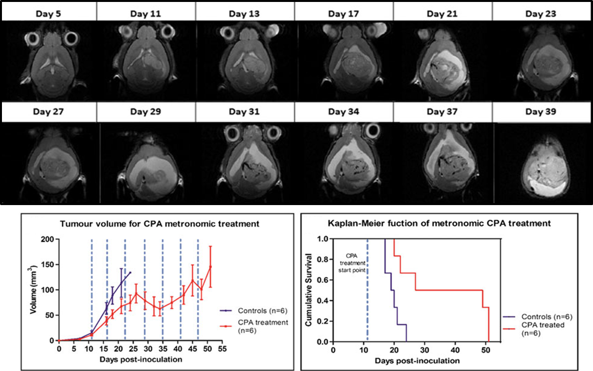
Glioblastoma (GBM) is the most common and aggressive glial primary tumor with a survival average of 14-15 months, even after application of standard treatment. Non-invasive surrogate biomarkers of therapy response may be relevant for improving patient survival. Nosological images of therapy response using a semi-supervised source extraction approach in preclinical GBM based on single slice Magnetic Resonance Spectroscopic Imaging (MRSI) was previously describe by our group. However, because of GBM heterogeneity, relevant response information could be missed just by studying one slice. 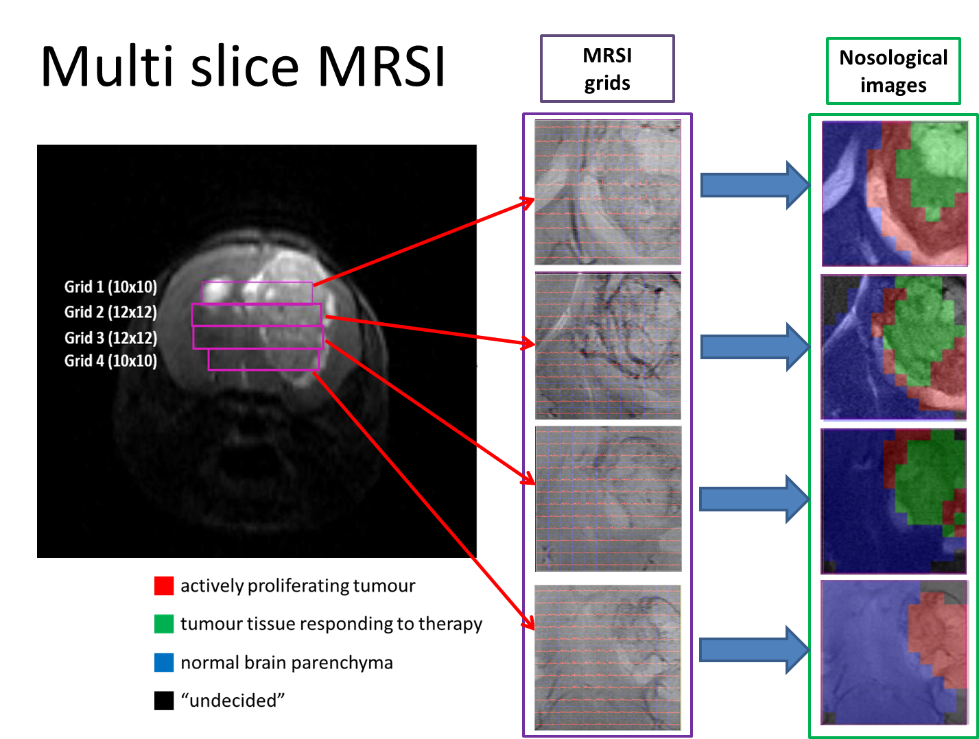 Therefore, the goal of this work was to acquire 3D-like information from preclinical GBM under a longitudinal treatment protocol, using a multi-slice MRSI approach.
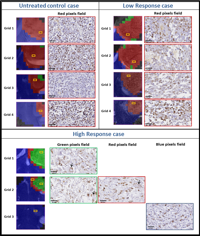
Nosological maps were obtained based on semi-supervised convex Non-negative Matrix Factorization and each voxel was colored according to the contribution to the spectral pattern of each one of the three sources or characteristic spectral patterns: Normal brain, actively proliferating tumour or responding tumour.
Heterogeneous response levels were observed and three arbitrary groups of treated animals were defined as: high response, intermediate response, and low response. Histopathological studies showed an inverse correlation between the responding pattern level and Ki67 proliferation rate.