Monthly Archives: October 2016

RDC measurements and application

joceah_v081i019.inddApplication to the structural discrimination of small molecules containing multiple stereocenters. One-Shot determination of residual dipolar couplings.
Castañar, L.; Garcia, M.; Hellemann, E.; Nolis, P.; Gil, R.; Parella, T.


A novel approach for the fast and efficient structural discrimination of molecules containing multiple stereochemical centers is described. A robust Jresolved HSQC experiment affording highly resolved 1JCH/1TCHsplittings along the indirect dimension and homodecoupled 1H signals in the detected dimension is proposed. The experiment enables in-situ distinction of both isotropic and anisotropic components of molecules dissolved incompressed PMMA gels, allowing a rapid and direct one-shot determination of accurate residual dipolar coupling constants from a single NMR spectrum


toc_joc_rdc

Access to experimentally infeasible spectra by pure-shift NMR covariance

Access to experimentally infeasible spectra by pure-shift NMR covariance. André Fredi, Pau Nolis, Carlos Cobas and Teodor Parella. Journal of Magnetic Resonance, Volume 270, September 2016, Pages 161-168. DOI: 10.1016/j.jmr.2016.07.010

Abstract: Covariance processing is a versatile processing tool to generate synthetic NMR spectral representations without the need to acquire time-consuming experimental datasets. Here we show that even experimentally prohibited NMR spectra can be reconstructed by introducing key features of a reference 1D CHn-edited spectrum into standard 2D spectra. This general procedure is illustrated with the calculation of experimentally infeasible multiplicity-edited pure-shift NMR spectra of some very popular homonuclear (ME-psCOSY and ME-psTOCSY) and heteronuclear (ME-psHSQC-TOCSY and ME-psHMBC) experiments.

Continue reading Access to experimentally infeasible spectra by pure-shift NMR covariance

10th Workshop on Magnetic Resonance Spectroscopy and Imaging (MRI/MRS) Applied to Laboratory Animals

Workshop dates: November 29th – December 2nd, 2016
Registration deadline: November 14th, 2016
Registration: REGISTRATION CLOSED  online
Capacity: Workshop limited to 4 participants (first come, first served)
Contact person: Silvia Lope-Piedrafita, PhD ()

This course combines a comprehensive series of lectures on the technology of Magnetic resonance spectroscopy and imaging (MRS/MRI) with hands-on laboratory sessions to provide practical demonstrations of key concepts and procedures for preclinical studies.

Whether you are considering MRI as a research tool in your lab or just would like to learn more about MRI, this workshop addresses practical aspects of experimental MRI with laboratory animals and provide valuable hands-on experience on a 7 Tesla Bruker BioSpec spectrometer.20161001_10cursomrimrs

See the workshop brochure for more information or contact Dra. Silvia Lope via email.

Mycobacteria clumping increase their capacity to damage macrophages

frontiersmicrobiology


“Mycobacteria clumping increase their capacity to damage macrophages” by C. Brambilla, M. Llorens-Fons, E. Julián, E. Noguera-Ortega, C. Tomàs-Martínez, M. Pérez-Trujillo, T. F. Byrd, F. Alcaide and M. Luquin.

Front. Microbiol. 7:1562.  DOI: 10.3389/fmicb.2016.01562

The rough morphotypes of non-tuberculous mycobacteria have been associated with the most severe illnesses in humans. This idea is consistent with the fact that Mycobacterium tuberculosis presents a stable rough morphotype. Unlike smooth morphotypes, the bacilli of rough morphotypes grow close together, leaving no spaces among them and forming large aggregates (clumps). Currently, the initial interaction of macrophages with clumps remains unclear. Thus, we infected J774 macrophages with bacterial suspensions of rough morphotypes of Mycobacterium abscessus containing clumps and suspensions of smooth morphotypes, primarily containing isolated bacilli. Using confocal laser scanning microscopy and electron microscopy, we observed clumps of at least 5 rough-morphotype bacilli inside the phagocytic vesicles of macrophages at 3 hours post-infection. These clumps grew within the phagocytic vesicles, killing 100% of the macrophages at 72 hours post-infection, whereas the proliferation of macrophages infected with smooth morphotypes remained unaltered at 96 hours post-infection. Thus, macrophages phagocytose large clumps, exceeding the bactericidal capacities of these cells. Furthermore, proinflammatory cytokines and granuloma-like structures were only produced by macrophages infected with rough morphotypes. Thus, the present study provides a foundation for further studies that consider mycobacterial clumps as virulence factors.

fig8

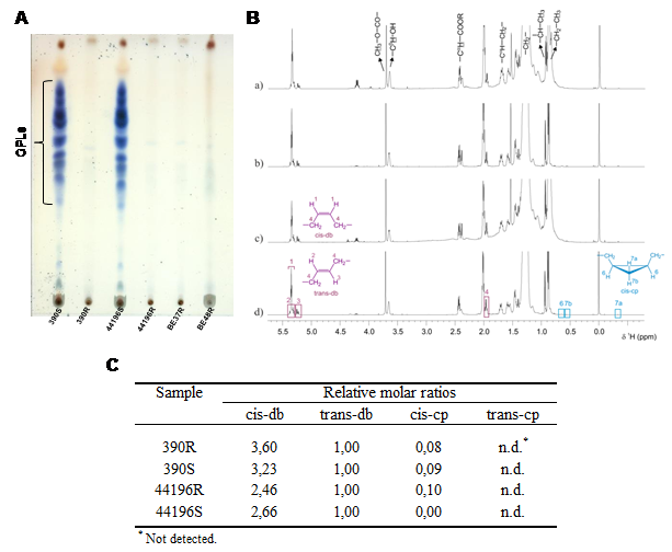
Figure. Content of GPL and structure of mycolic acids. (A) 1-D TLC analysis of the crude lipid extracts of M. abscessus strains. (B) 1H-NMR spectra of purified mycolic acid methyl esters from M. abscessus. (C) Relative molar ratios of molecular moieties cis-db, trans-db, cis-cp and trans-cp of mycolic acid methyl esters from M. abscessus.
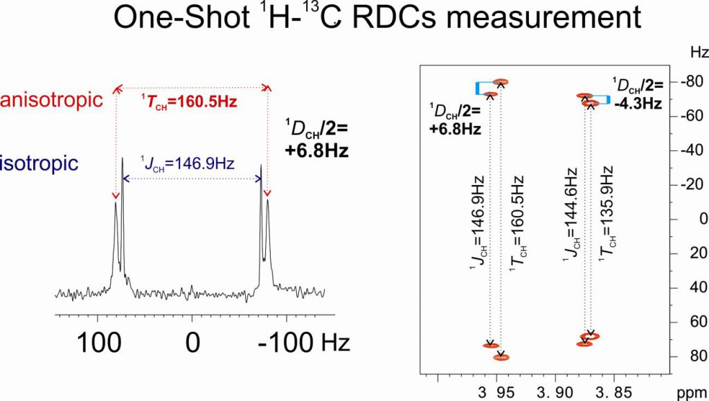