Tag Archives: MRSI

Multi-Slice MRSI Analysis of Therapy Response in Preclinical Glioblastoma

Metabolomics of Therapy Response in Preclinical Glioblastoma: A Multi-Slice MRSI-Based Volumetric Analysis for Noninvasive Assessment of Temozolomide Treatment” by N. Arias-Ramos, L. Ferrer-Font,  S. Lope-Piedrafita,  V. Mocioiu, M. Julià-Sapé , M. Pumarola, C. Arús  and A. P. Candiota. Metabolites, 2017, 18;7(2). pii: E20. DOI: 10.3390/metabo7020020.

Glioblastoma (GBM) is the most common and aggressive glial primary tumor with a survival average of 14-15 months, even after application of standard treatment. Non-invasive surrogate biomarkers of therapy response may be relevant for improving patient survival. Nosological images of therapy response using a semi-supervised source extraction approach in preclinical GBM based on single slice Magnetic Resonance Spectroscopic Imaging (MRSI) was previously describe by our group. However, because of GBM heterogeneity, relevant response information could be missed just by studying one slice. Therefore, the goal of this work was to acquire 3D-like information from preclinical GBM under a longitudinal treatment protocol, using a multi-slice MRSI approach.
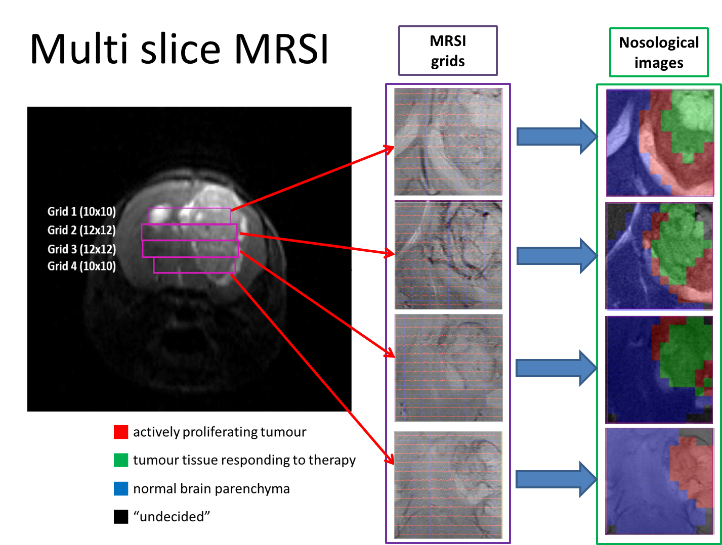
Nosological maps were obtained based on semi-supervised convex Non-negative Matrix Factorization and each voxel was colored according to the contribution to the spectral pattern of each one of the three sources or characteristic spectral patterns: Normal brain, actively proliferating tumour or responding tumour.

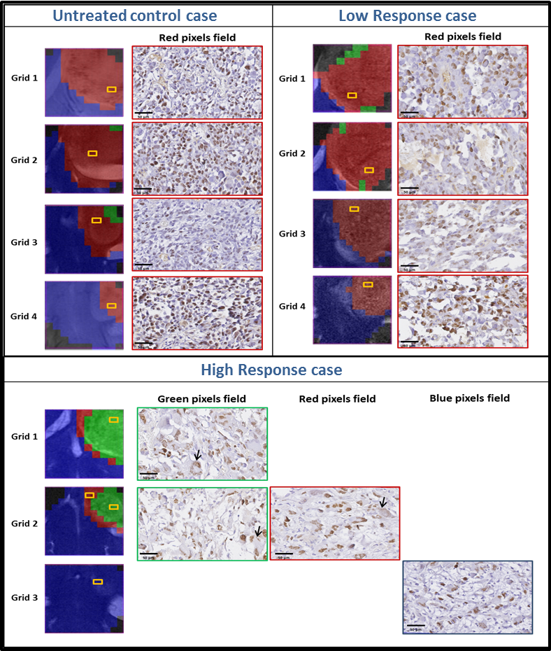
Heterogeneous response levels were observed and three arbitrary groups of treated animals were defined as: high response, intermediate response, and low response. Histopathological studies showed an inverse correlation between the responding pattern level and Ki67 proliferation rate.
 

Job offer for the TRANSACT-ITN project

We are recruiting an Early Stage Researcher to work on the Development of Decision Support System & Spectral Classification tool meta-plug-ins for the jMRUI platform as part of the TRANSACT-ITN European project.

QR-code of ESR-UAB job offer at EURAXESS site
Scan the QR-code to get this job offer link at the EURAXESS Jobs Portal.

We seek a highly motivated and qualified individual as Early Stage Researcher for a three-year applied research project. The successful candidate will contribute to the development of advanced biomedical research tools in the field of Magnetic Resonance Spectroscopy and Imaging, and its application to the clinical day-to-day practice.

The position is intended for a young post-graduate student/researcher, with a Master degree or equivalent on Engineering or Computer Science, and with a proficient knowledge of the Java programming language. Prior experience on signal and/or image processing, segmentation and pattern recognition applications in the field of biomedical research is an asset, particularly in the field of Magnetic Resonance Spectroscopy and/or Imaging.

Continue reading Job offer for the TRANSACT-ITN project

TRANSACT-ITN project awarded to GABRMN, SeRMN & CIBER-BBN

TRANSCAT ITN ProjectTransforming Magnetic Resonance Spectroscopy into a Clinical Tool (TRANSACT) is an European research project awarded in the call FP7-PEOPLE-2011-ITN, of the 7th Framework Programme Marie-Curie Actions, to a network of partners including the GABRMN and SeRMN at UAB, and the associated partner CIBER-BBN. The project is coordinated by Prof. Sabine Van Huffel, Prof. Uwe Himmelreich, and Dr. Diana Sima, of the Department of Electrical Engineering ESAT-SCD, and Department of Imaging & Pathology, Biomedical MRI Unit, Katholieke Universiteit Leuven, Leuven, Belgium. Scientist-in-charge at UAB will be Prof. Carles Arús (GABRMN) and Miquel Cabañas (SeRMN), and Dr. Margarida Julià-Sapé will be scientist-in-charge at CIBER-BBN. Dr. Silvia Lope-Piedrafita (SeRMN) will take part in the project as senior scientist.

Continue reading TRANSACT-ITN project awarded to GABRMN, SeRMN & CIBER-BBN

DMSO as a magnetic resonance contrast agent for brain tumors

“Dimethyl sulfoxide (DMSO) as a potential contrast agent for brain tumors”, by Delgado-Goñi T, Martín-Sitjar J, Simões RV, Acosta M, Lope-Piedrafita S, Arús C.; NMR in Biomedicine. 2012.DOI: 10.1002/nbm.2832.

Identifying the type of brain tumor helps doctors determine the most appropriate course of treatment. Brain tumor diagnosis usually involves a neurological examination, brain scans, and/or an analysis of the brain tissue. Nowadays, the biopsy, although it is a very invasive procedure, is the most accurate method of obtaining a diagnosis. Continue reading DMSO as a magnetic resonance contrast agent for brain tumors