Workshop limited to 4 participants (first come, first served)
Contact person:
Silvia Lope-Piedrafita, PhD ()
This course combines a comprehensive series of lectures on the technology of Magnetic resonance spectroscopy and imaging (MRS/MRI) with hands-on laboratory sessions to provide practical demonstrations of key concepts and procedures for preclinical studies.
Whether you are considering MRI as a research tool in your lab or just would like to learn more about MRI, this workshop addresses practical aspects of experimental MRI with laboratory animals and provide valuable hands-on experience on a 7 Tesla Bruker BioSpec spectrometer.
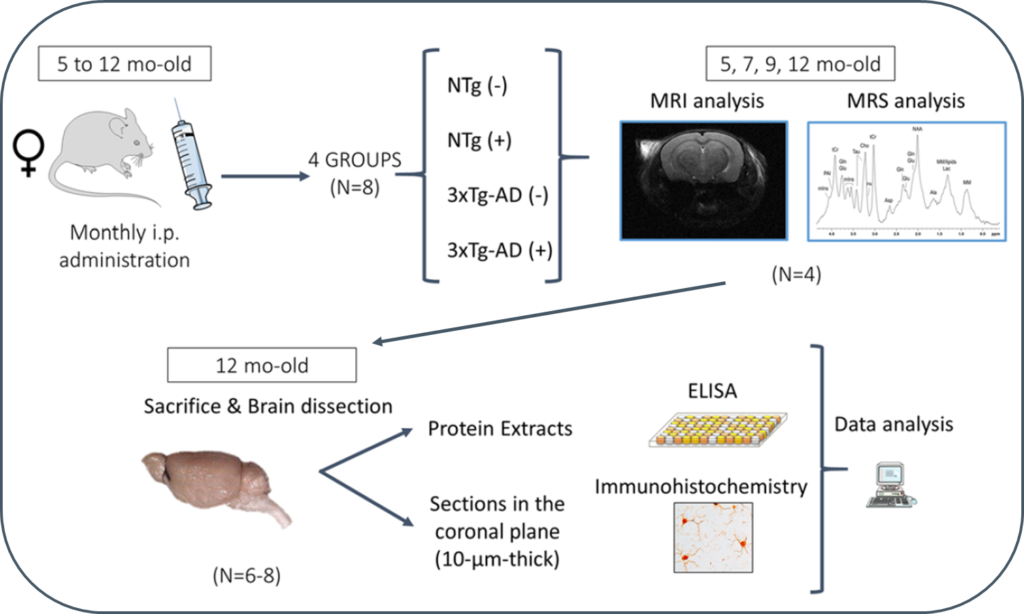
Progression of Alzheimer’s disease and effect of scFv-h3D6 immunotherapy in the 3xTg-AD mouse model: An in vivo longitudinal study using Magnetic Resonance Imaging and Spectroscopy by Güell-Bosch J, Lope-Piedrafita S, Esquerda-Canals G, Montoliu-Gaya L, and Villegas S. NMR in Biomedicine 33(5):e4263; DOI: 10.1002/nbm.4263.
Alzheimer’s disease (AD) is an incurable disease that affects most of the 47 million people estimated as living with dementia worldwide. The main histopathological hallmarks of AD are extracellular β-amyloid (Aβ) plaques and intracellular neurofibrillary tangles (NFTs) composed of hyperphosphorylated tau protein. In recent years, Aβ-immunotherapy has been revealed as a potential tool in AD treatment. One strategy consists of using single-chain variable fragments (scFvs), which avoids the fragment crystallizable (Fc) effects that are supposed to trigger a microglial response, leading to microhemorrhages and vasogenic edemas, as evidenced in clinical trials with bapineuzumab. The scFv-h3D6 generated by our research group derives from this monoclonal antibody, which targets the N-terminal of the Aβ peptide and recognizes monomers, oligomers and fibrils.
In this study, 3xTg-AD mice were intraperitoneally and monthly treated with 100 μg of scFv-h3D6 (a dose of ~3.3 mg/kg) or PBS, from 5 to 12 months of age (-mo), the age at which the mice were sacrificed and samples collected for histological and biochemical analyses. During treatments, four monitoring sessions using magnetic resonance imaging and spectroscopy (MRI/MRS) were performed at 5, 7, 9, and 12 months of age. MRI/MRS techniques allow, in a non-invasive manner, to draw an in vivo picture of concrete aspects of the pathology and to monitor its development across time. Compared with the genetic background, 3xTg-AD mice presented a smaller volume in almost all cerebral regions and ages examined, an increase in both the intra and extracellular Aβ1-42 at 12-mo, and an inflammation process at this age, in both the hippocampus (IL-6 and mIns) and cortex (IL-6). In addition, treatment with scFv-h3D6 partially recovered the values in brain volume, and Aβ, IL-6, and mIns concentrations, among others, encouraging further studies with this antibody fragment.
“Neonatal handling enduringly decreases anxiety and stress responses and reduces hippocampus and amygdala volume in agenetic model of differential anxiety: Behavioral-volumetric associations in the Roman rats trains” by C. Río-Álamos, I. Oliveras, M. A. Piludu, C. Gerbolés, T. Cañete, G. Blázquez, S. Lope-Piedrafita, E. Martínez-Membrives, R. Torrubia, A. Tobeña, and A. Fernández-Teruel. European Neuropsychopharmacology, 2017, 27: 146–158. DOI: 10.1016/j.euroneuro.2016.12.003
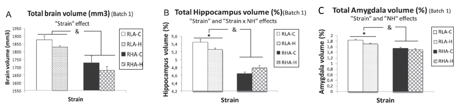
The hippocampus and amygdala have been proposed as key neural structures related to anxiety. A more active hippocampus/amygdala system has been related to greater anxious responses in situations involving conflict/novelty. The Roman Low- (RLA) and High-avoidance (RHA) rat strains constitute a genetic model of differential anxiety. Relative to RHA rats, RLA rats exhibit enhanced anxiety/fearfulness, augmented hippocampal/amygdala c-Fos expression following exposure to novelty/conflict, increased hippocampal neuronal density and higher endocrine responses to stress. Neonatal handling (NH) is an environmental treatment with long-lasting anxiety/stress-reducing effects in rodents. Since hippocampus and amygdala volume are supposed to be related to anxiety/fear, it was hypothesized a greater volume of both areas in RLA than in RHA rats, as well as that NH treatment would reduce anxiety and the volume of both structures. Adult untreated and NH-treated RHA and RLA rats were tested for anxiety, sensorimotor gating (PPI), stress-induced corticosterone and prolactin responses, two-way active avoidance acquisition and in vivo 7 T 1H-Magnetic resonance image.
As expected, untreated RLA rats showed higher anxiety and post-stress hormone responses, as well as greater hippocampus and amygdala volumes than untreated RHA rats. NH decreased anxiety/stress responses, especially in RLA rats, and significantly reduced hippocampus and amygdala volumes in this strain. Dorsal striatum volume was not different between the strains nor it was affected by NH. Finally, there were positive associations (as shown by correlations, factor analysis and multiple regression) between anxiety and PPI and hippocampus/amygdala volumes.
 “Neonatal handling enduringly decreases anxiety and stress responses and reduces hippocampus and amygdala volume in agenetic model of differential anxiety: Behavioral-volumetric associations in the Roman rats trains” by C. Río-Álamos, I. Oliveras, M. A. Piludu, C. Gerbolés, T. Cañete, G. Blázquez,