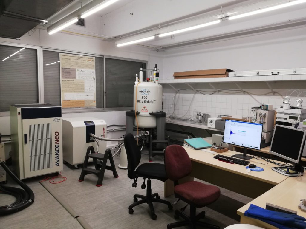
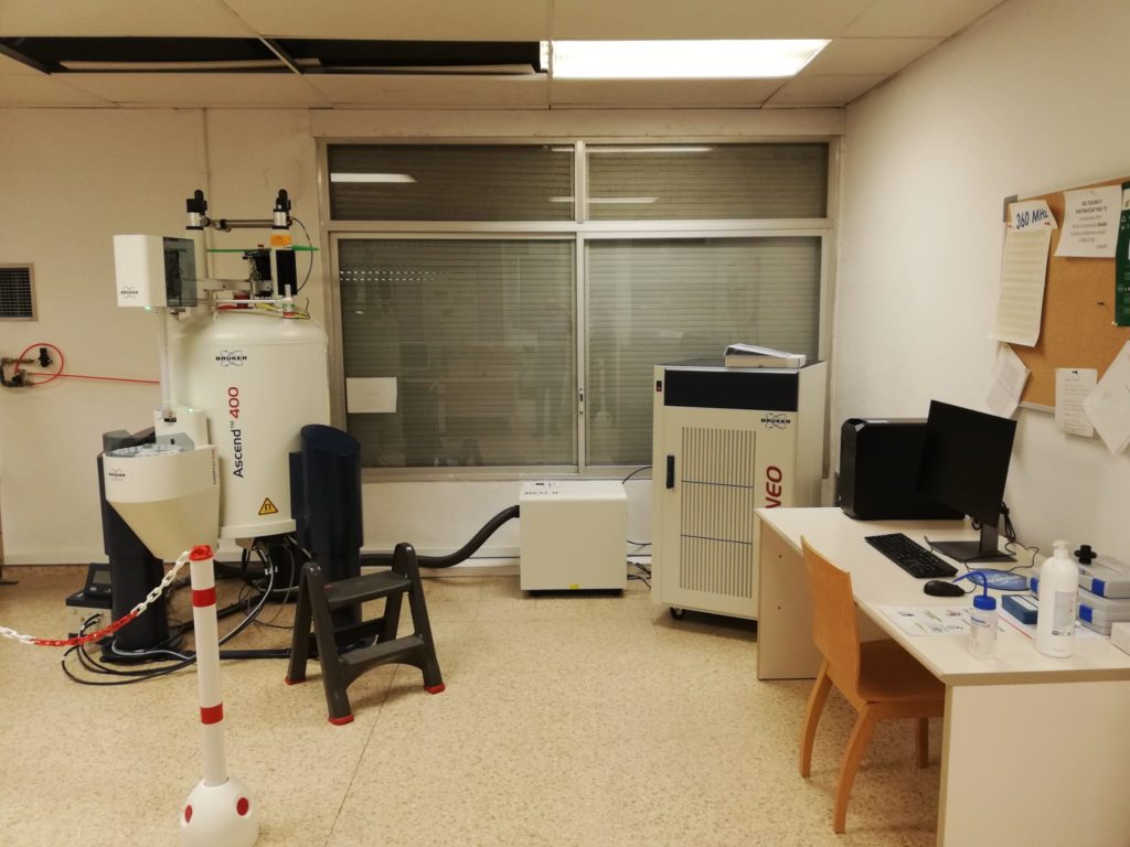
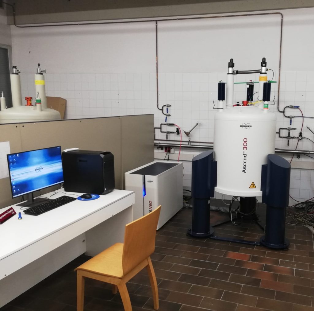
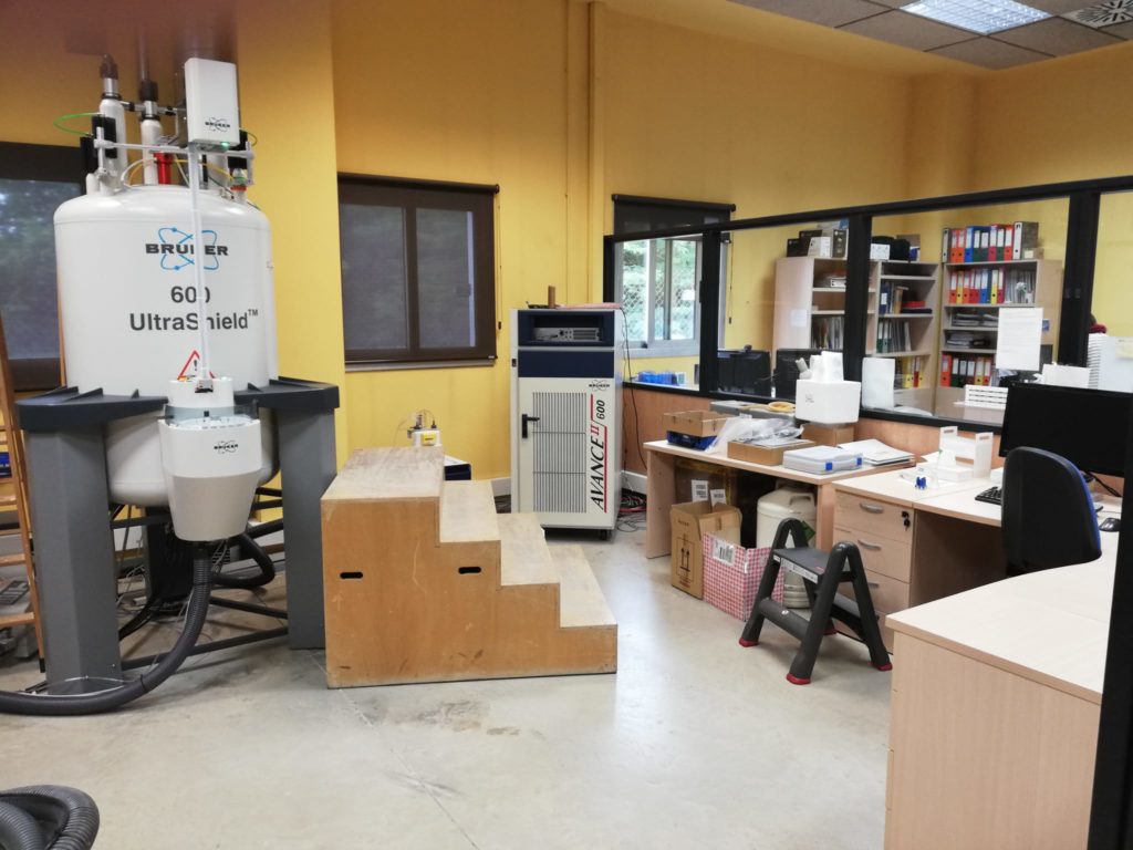
To improve the obsolescence of the oldest equipments, the following new NMR spectrometers have been installed/updated at the SeRMN-UAB Facility in May 2021:
A new AVANCE NEO console for the existing 500 MHz NMR spectrometer, equipped with an automated ATMA accessory and a compatible cryoplatform for the current TCI cryoprobe.
A New 400 MHz NMR spectrometer including a ultrashielded ASCEND magnet, automated SAMPLE-CASE sample changer, high-resolution liquid (iProbe) and solid-state (CP-MAS 4.0mm) NMR probes, and a BCU-II unit for automated sample refrigeration until 233K.
3. A New 300 MHz NMR spectrometer including a Nanobay AVANCE nanoNEO console, ultra shielded ASCEND magnet and a BBFO probe head.
4. A cooled SAMPLE-CASE sample changer and a BCU-II unit for automated sample refrigeration until 233K for the existing 600MHz NMR spectrometer
More info about the equipments and accesories can be found at the Bruker WWW site.
The purchase of these equipments has been co-financed by the Ministry of Science, Innovation, and Universities to the 2019 infrastructure call (project EQC2019-005396-P) co-financed by the European Fund for Economic and Regional Development (FEDER) through the plurirregional operating program of Spain (POPE) period 2014-2020.
From 22-6-2020, all authorized SeRMN users can make use of the self-service mode in the 250auto, 360MHz and 400MHz spectrometers, exclusively from 9AM to 5PM, using the booking program (https://sermn.uab.cat/reserves/). The experiment request service is also active for all those samples that are not recorded in self-service mode:
At the moment, the 250robot spectrometer is reserved exclusively for this type of work.
It is very important that the following mandatory rules are respectedt 1. Self-service exclusively from 9AM to 5PM. SeRMN access is not allowed outside of this time slot as there will be no SeRMN staff. 2. Only 1 person per machine is allowed. Always respect two meters of distance separation between people. 3. It is necessary to follow the established protocols of hygiene and safety at a personal level (mask, hand disinfection …) 4. Before and, above all, after using the keyboard and other tools to carry out the experiments (spinner, calibrator …) it is necessary to disinfect them with the hygiene material that you will find available.
Any questions or clarifications, you can contact the staff of the SeRMN who will be present from 9AM to 5PM or through the address .
Després de més de dos mesos d’obligat tancament, el SeRMN reobrirà les seves instal.lacions a partir del proper dilluns dia 25 de Maig. Ja que s’han de mantenir certes precaucions de seguretat i higiene, començarem amb un funcionament limitat, provisional i progressiu a mida que es vagi normalitzant la situació del COVID19. Les normes bàsiques de funcionament són:
1. No hi ha autoservei. No cal fer reserva en el nostre sistema de reserves com es fa habitualment. Les mostres seran analitzades exclusivament pel personal del SeRMN.
2. Prèviament, cal fer una sol.licitud de prestació de servei per a cada mostra a través dels formularis de la nostra plana web:
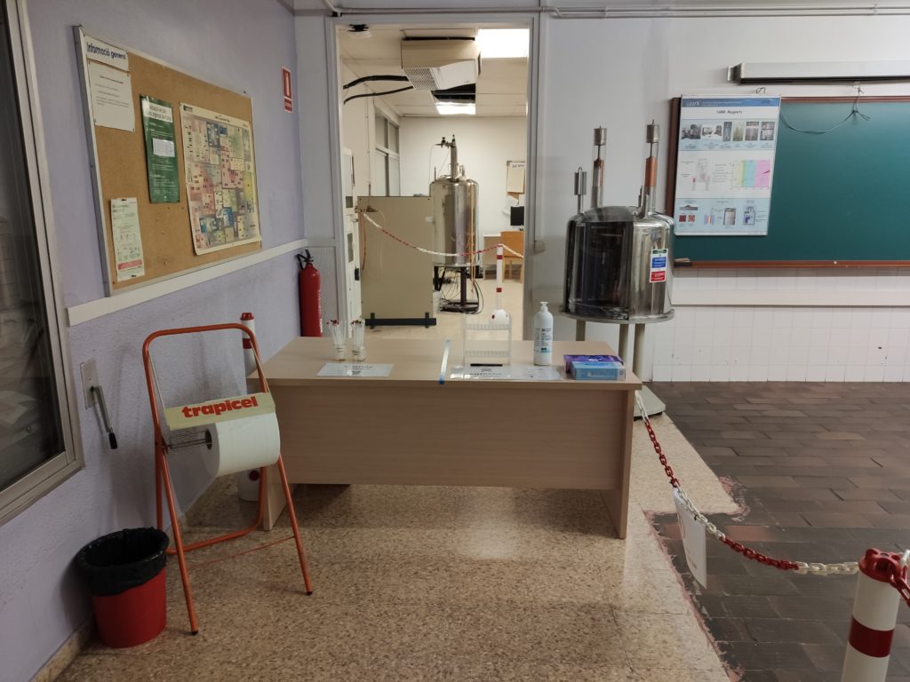
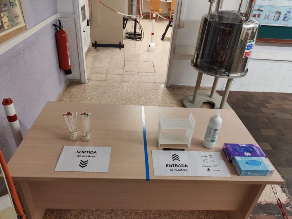
3. Pels usuaris que treballin a l’edifici de les Facultats de Ciències i Biociències, cal portar la mostra presencialment al SeRMN en horari de matí (9AM-1PM). És important que la mostra estigui ben etiquetada i prèviament desinfectada. L’accés al SeRMN està restringit. Just a l’entrada del SeRMN hi haurà una taula de recepció per desinfectar/deixar/recuperar les mostres (veure https://sct.uab.cat/sermn/sample_delivery_provisional).
4. Degut a l’accés restringit que hi ha a l’edifici de les Facultats de Ciències i Biociències, recomanen als usuaris externs sense autorització d’accés a l’edifici (altres facultats de la UAB, instituts i empreses del PRUAB, i instituts i empreses externes al PRUAB) que es posin en contacte amb el personal del SeRMN per email o per telèfon (93 581 3785 o al 93 581 2291) per concretar el lliurament/recollida de mostres.
Per qualsevol dubte o qüestió, podeu contactar-nos a través del nostre mail institucional () o en els nostres correus personals.
————————————————————————————————————————–
After more than two months of forced closure, the SeRMN will reopen its facilities from next Monday, May 25. Since certain safety and hygiene precautions must be maintained, we will start with a limited, provisional and progressive operation as the COVID-19 situation normalizes. The basic rules of operation are:
1-. There is no self-service. There is no need to make a reservation in our booking system as usual. Samples will be analyzed exclusively by the SeRMN staff.
2-. Previously, a request for service for each sample through the forms on our website is required:
3.- For users working in the building of the Faculties of Sciences and Biosciences, the sample must be brought in person at the SeRMN in the morning (9 AM-1PM). It is important that the sample is well labeled and previously disinfected. The access to the SeRMN lab is restricted. Right at the entrance of the SeRMN there will be a reception table where you must disinfect / leave / recover your samples (see https://sct.uab.cat/sermn/sample_delivery_provisional).
4. Due to the restricted access to the building of the Faculties of Sciences and Biosciences, we strongly recommend to external users without authorization access to the building (other faculties of the UAB, institutes and companies of the PRUAB, and institutes and companies external to the PRUAB) that contact with the staff of the SeRMN by email or telephone (93 581 3785 or 93 581 2291) to specify the delivery of samples.
For any questions or concerns, you can contact us via our institutional email () or in our personal emails.
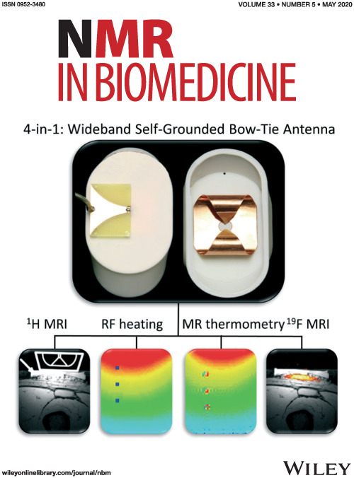
Progression of Alzheimer’s disease and effect of scFv-h3D6 immunotherapy in the 3xTg-AD mouse model: An in vivo longitudinal study using Magnetic Resonance Imaging and Spectroscopy by Güell-Bosch J, Lope-Piedrafita S, Esquerda-Canals G, Montoliu-Gaya L, and Villegas S. NMR in Biomedicine 33(5):e4263; DOI: 10.1002/nbm.4263.
Alzheimer’s disease (AD) is an incurable disease that affects most of the 47 million people estimated as living with dementia worldwide. The main histopathological hallmarks of AD are extracellular β-amyloid (Aβ) plaques and intracellular neurofibrillary tangles (NFTs) composed of hyperphosphorylated tau protein. In recent years, Aβ-immunotherapy has been revealed as a potential tool in AD treatment. One strategy consists of using single-chain variable fragments (scFvs), which avoids the fragment crystallizable (Fc) effects that are supposed to trigger a microglial response, leading to microhemorrhages and vasogenic edemas, as evidenced in clinical trials with bapineuzumab. The scFv-h3D6 generated by our research group derives from this monoclonal antibody, which targets the N-terminal of the Aβ peptide and recognizes monomers, oligomers and fibrils.
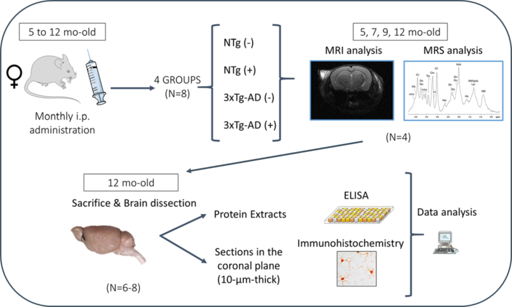
In this study, 3xTg-AD mice were intraperitoneally and monthly treated with 100 μg of scFv-h3D6 (a dose of ~3.3 mg/kg) or PBS, from 5 to 12 months of age (-mo), the age at which the mice were sacrificed and samples collected for histological and biochemical analyses. During treatments, four monitoring sessions using magnetic resonance imaging and spectroscopy (MRI/MRS) were performed at 5, 7, 9, and 12 months of age. MRI/MRS techniques allow, in a non-invasive manner, to draw an in vivo picture of concrete aspects of the pathology and to monitor its development across time. Compared with the genetic background, 3xTg-AD mice presented a smaller volume in almost all cerebral regions and ages examined, an increase in both the intra and extracellular Aβ1-42 at 12-mo, and an inflammation process at this age, in both the hippocampus (IL-6 and mIns) and cortex (IL-6). In addition, treatment with scFv-h3D6 partially recovered the values in brain volume, and Aβ, IL-6, and mIns concentrations, among others, encouraging further studies with this antibody fragment.
Due to the alarm status by the Covid-19 the UAB’s Nuclear Magnetic Resonance Service will be temporally closed. For any questions you can contact us by email at the institutional address
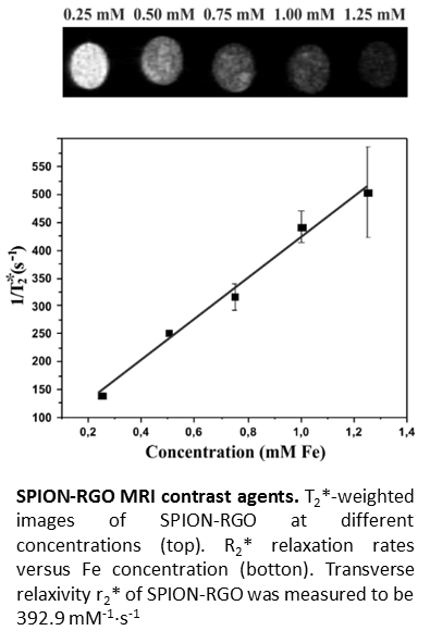
Magnetic resonance imaging (MRI) is a useful tool for disease diagnosis
and treatment monitoring. Superparamagnetic iron oxide nanoparticles (SPION)
show good performance as transverse relaxation (T2) contrast agents, thus
facilitating the interpretation of the acquired images. Attachment of SPION
onto nanocarriers prevents their agglomeration, improving the circulation time
and efficiency. Graphene derivatives, such as graphene oxide (GO) and reduced
graphene oxide (RGO), are appealing nanocarriers since they have both high
surface area and functional moieties that make them ideal substrates for the
attachment of nanoparticles. A fast, simple, and environmentally friendly
microwave-assisted approach for the synthesis of SPION-RGO hybrids has been demonstrated
in this study. Different iron precursor/GO ratios were used leading to SPION,
with a median diameter of 7.1 nm, homogeneously distributed along the RGO
surface. Good relaxivity (r2*) values were obtained in MRI studies and no
significant toxicity was detected within in vitro tests following GL261 glioma
and J774 macrophage-like cells for 24 h with SPION-RGO, demonstrating the
applicability of the hybrids as T2-weighted MRI contrast agents.
This is the first report on the obtention of functionalized MSN by a co-condensation procedure with a structurally complex chiral precursor. The functionalized MSN have been characterized by elemental analysis, 29Si and 13C CP MAS NMR, transmission electron microscopy, scanning electron microscopy, N2-sorption measurements, dynamic light scattering, ζ-potential, and powder X-ray diffraction. We have evaluated the activity of these materials as recyclable catalysts in the asymmetric aldol reaction. The use of organosilica nanoparticles reduces the problems of diffusion and low reaction rates encountered with bulk organosilicas.
We are recruiting an Early Stage Researcher to work on a decision-support system based on MRSI data at 3T, for glioblastoma therapy response follow- up, as part of the INSPiRE-MED European project.
We seek a highly motivated and qualified individual as Early Stage Researcher for a three-year applied research project. The successful candidate will contribute to the development of advanced biomedical research tools in the field of Magnetic Resonance Spectroscopy and Imaging, and its application to the clinical day-to-day practice.
Project description: This position is one of the 15 ESR positions of the INSPiRE-MED European Training Network, which focuses on the development of Magnetic Resonance Spectroscopy (MRS) and MR Spectroscopic Imaging (MRSI) combined with Positron Emission Tomography (PET), enhanced by machine learning techniques.
The main aim of the PhD project (ESR12) will be development of a Machine Learning medical decision-support system based on MRSI data at 3T, for glioblastoma therapy response follow-up.
The
ESR will develop a novel medical decision support system (MDSS)
focused on glioblastoma therapy response follow-up, based on magnetic
resonance spectroscopic imaging (MRSI) data, able to take and process
data from multiple MRSI formats and centres. For each patient’s
MRSI, the MDSS should deliver a nosological or classification image,
ready to be fused with images of other MR modalities from the same
patient. The DSS will be integrated into the interface of the
academic version of jMRUI, in a way that allows clinicians evaluate
the system with their data. An important part of of the project will
be the incorporation of automated MRSI artifact detection and removal
tools.
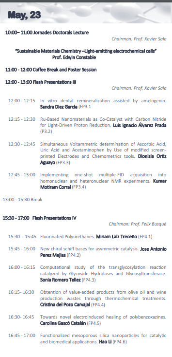
The 23rd May 2019 at 12:45, Sala d’Actes de la Facultat de Ciències de la UAB, I will present my work on “Implementing one-shot multiple-FID acquisition into homonuclear and heteronuclear NMR experiments” at the Novena Edició de les Jornades Doctorals by the PhD Chemistry Program and the Chemistry Department (Meeting program).
Official call by Universitat Autònoma de Barcelona
Deadline for submissions: 21/5/2019 at 23:00
See UAB and/or Euraxess advertisements for further information about the position and how to apply.
We are recruiting an Early Stage Researcher to work on the implementation of high-resolution MRSI methods in a pre-clinical scanner as part of the INSPiRE-MED European project.
We seek a highly motivated and qualified individual as Early Stage Researcher for a three-year applied research project. The successful candidate will contribute to the development of advanced biomedical research tools in the field of Magnetic Resonance Spectroscopy and Imaging, and its application to the clinical day-to-day practice.
Project description: This position is one of the 15 ESR positions of the INSPiRE-MED European Training Network, which focuses on the development of Magnetic Resonance Spectroscopy (MRS) and MR Spectroscopic Imaging (MRSI) combined with Positron Emission Tomography (PET), enhanced by machine learning techniques.
The main aim of the PhD project (ESR4) will be the implementation of innovative high spatial resolution MRSI methods in a pre-clinical scanner. The ultimate goal will be the validation of optimal methods for improving imaging biomarker development of brain tumour in longitudinal studies of therapy response in mouse glioblastoma models. The project will involve evaluation of the methodology performance limits, repeatability and reproducibility compared to stock Bruker Biospec MRSI sequences and the assessment of speed-up MRSI methods in a 7-Tesla pre-clinical scanner.

 by Llenas M, Sandoval S, Costa PM, Oró-Solé J,