Tag Archives: MRI

related to mr imaging.

8th Workshop on Magnetic Resonance Spectroscopy and Imaging (MRI/MRS) Applied to Laboratory Animals

Dates:  10th to 13th June 2013

Organized by the SeRMN of the Autonomous University of Barcelona (UAB).
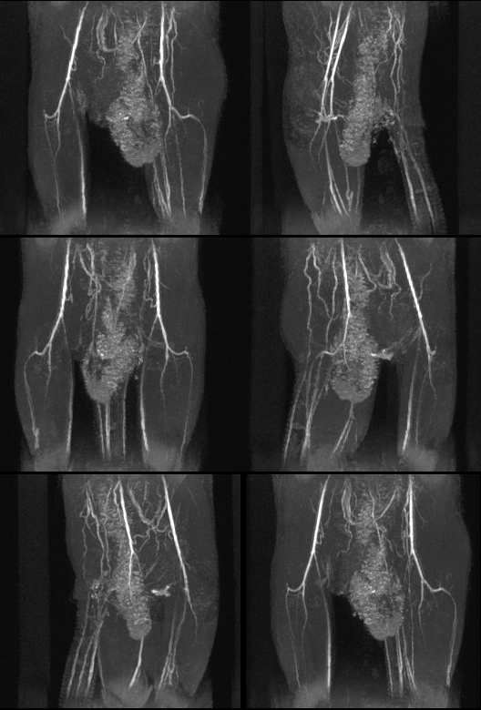
LimbAngio_3DTOFThis workshop combines a comprehensive series of lectures on the technology of Magnetic resonance spectroscopy and imaging (MRS/MRI) with hands-on laboratory sessions to provide practical demonstrations of key concepts and procedures for preclinical studies.

Whether you are considering MRI as a research tool in your lab or just would like to learn more about MRI, this workshop addresses practical aspects of experimental MRI with laboratory animals and provide valuable hands-on experience on a 7 Tesla Bruker BioSpec spectrometer.

Number of participants will be limited to 4.

For the registration, please fill the Registration Form and email it to (registration deadline ends May 27st)

See Workshop Brochure for more information or contact Dra. Silvia Lope.


Brain magnetic resonance in liver failure

“Brain magnetic resonance in experimental acute-on-chronic liver failure” by L. Chavarria, M. Oria, J. Romero-Giménez, J. Alonso, S. Lope-Piedrafita, and J. Cordoba.
Liver International 33 (2013) 294-300. DOI: 10.1111/liv.12032

Liver failure causes brain edema that can lead to intracranial hypertension and death. It is currently uncertain whether this water accumulation is predominantly intracellular or extracellular, something important to determine in order to apply the most adequate therapeutic measures. The aim of this study was to investigate the mechanisms involved in the neurological manifestations occurring in an experimental acute-on-chronic liver failure rat model that combines sustained liver injury with a factor inducing an inflammatory response.

Continue reading Brain magnetic resonance in liver failure

Job offer for the TRANSACT-ITN project

We are recruiting an Early Stage Researcher to work on the Development of Decision Support System & Spectral Classification tool meta-plug-ins for the jMRUI platform as part of the TRANSACT-ITN European project.

QR-code of ESR-UAB job offer at EURAXESS site
Scan the QR-code to get this job offer link at the EURAXESS Jobs Portal.

We seek a highly motivated and qualified individual as Early Stage Researcher for a three-year applied research project. The successful candidate will contribute to the development of advanced biomedical research tools in the field of Magnetic Resonance Spectroscopy and Imaging, and its application to the clinical day-to-day practice.

The position is intended for a young post-graduate student/researcher, with a Master degree or equivalent on Engineering or Computer Science, and with a proficient knowledge of the Java programming language. Prior experience on signal and/or image processing, segmentation and pattern recognition applications in the field of biomedical research is an asset, particularly in the field of Magnetic Resonance Spectroscopy and/or Imaging.

Continue reading Job offer for the TRANSACT-ITN project

TRANSACT-ITN project awarded to GABRMN, SeRMN & CIBER-BBN

TRANSCAT ITN ProjectTransforming Magnetic Resonance Spectroscopy into a Clinical Tool (TRANSACT) is an European research project awarded in the call FP7-PEOPLE-2011-ITN, of the 7th Framework Programme Marie-Curie Actions, to a network of partners including the GABRMN and SeRMN at UAB, and the associated partner CIBER-BBN. The project is coordinated by Prof. Sabine Van Huffel, Prof. Uwe Himmelreich, and Dr. Diana Sima, of the Department of Electrical Engineering ESAT-SCD, and Department of Imaging & Pathology, Biomedical MRI Unit, Katholieke Universiteit Leuven, Leuven, Belgium. Scientist-in-charge at UAB will be Prof. Carles Arús (GABRMN) and Miquel Cabañas (SeRMN), and Dr. Margarida Julià-Sapé will be scientist-in-charge at CIBER-BBN. Dr. Silvia Lope-Piedrafita (SeRMN) will take part in the project as senior scientist.

Continue reading TRANSACT-ITN project awarded to GABRMN, SeRMN & CIBER-BBN

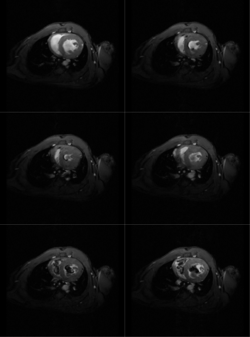
Cardiac MRI of the Sirt1-deficient mouse heart

“Dilated cardiomyopathy and mitochondrial dysfunction in Sirt1-deficient mice: A role for Sirt1-Mef2 in adult heart”
by A. Planavila, E. Dominguez, M. Navarro, M. Vinciguerra, R. Iglesias, M. Giralt, S. Lope-Piedrafita, J. Ruberte, F. Villarroya. Journal of Molecular and Cellular Cardiology 53 (2012) 521-531. DOI: 10.1016/j.yjmcc.2012.07.019

The protein deacetylase Sirtuin-1 (Sirt1) is involved in the cardiac hypertrophic responses and cardiac embryo morphogenesis. However, the physiological function of Sirt1 deficiency in the postnatal development of the heart remains to be characterized. The aim of this study was to investigate the relevance of Sirt1 in the development and function of the myocardium by using complementary techniques, such as gene expression, immunoblotting, immunohistochemistry, histological and electron microscopy examinations, and in vivo cardiac magnetic resonance imaging (MRI).

Continue reading Cardiac MRI of the Sirt1-deficient mouse heart

DMSO as a magnetic resonance contrast agent for brain tumors

“Dimethyl sulfoxide (DMSO) as a potential contrast agent for brain tumors”, by Delgado-Goñi T, Martín-Sitjar J, Simões RV, Acosta M, Lope-Piedrafita S, Arús C.; NMR in Biomedicine. 2012.DOI: 10.1002/nbm.2832.

Identifying the type of brain tumor helps doctors determine the most appropriate course of treatment. Brain tumor diagnosis usually involves a neurological examination, brain scans, and/or an analysis of the brain tissue. Nowadays, the biopsy, although it is a very invasive procedure, is the most accurate method of obtaining a diagnosis. Continue reading DMSO as a magnetic resonance contrast agent for brain tumors

Novel MRI contrast agents: peptide-SPION conjugates

Journal Cover:Chem. Commun., 2012, 48, 5322-5324“Efficient γ-amino-proline-derived cell penetrating peptide-superparamagnetic iron oxide nanoparticle conjugates via aniline-catalyzed oxime chemistry as bimodal imaging nanoagents”, by Cavalli S, Carbajo D, Acosta M, Lope-Piedrafita S, Candiota AP, Arús C, Royo M, Albericio F; Chem. Commun 48 (2012) p. 5322. DOI: 10.1039/C2CC17937G

Recent advances in nanotechnology have offered new opportunities for detection, prevention, and treatment of different diseases. In this respect engineered superparamagnetic iron oxide nanoparticles (SPIONs) represent an advanced tool in nanomedicine because they can be loaded in a multiple and orthogonal way with drugs and probes in order to simultaneously act as molecular imaging agents and drug carriers. Continue reading Novel MRI contrast agents: peptide-SPION conjugates

Neurodegeneration in NPC Mice by T2 and DTI MRI

“In Vivo Assessment of Neurodegeneration in Niemann-Pick Type C Mice by Quantitative T2 Mapping and Diffusion Tensor Imaging”, by John W. Totenhagen, Silvia Lope-Piedrafita, Ivan A. Borbon, Eriko S. Yoshimaru, Robert P. Erickson, and Theodore P. Trouard; Journal of Magnetic Resonance Imaging 35 (2012), 528-536. doi: 10.1002/jmri.22837

NIEMANN-PICK TYPE C (NPC) disease is a rare genetic neurodegenerative disease with no cure and few effective options for treatment. Diagnosis is most often made during childhood with symptoms progressively worsening and leading to death prior to adulthood. Continue reading Neurodegeneration in NPC Mice by T2 and DTI MRI

7th Workshop on Magnetic Resonance Spectroscopy and Imaging (MRI/MRS) Applied to Laboratory Animals

Dates:  24th -27th October 2011

Organized by the SeRMN of the Autonomous University of Barcelona (UAB).

This workshop combines a comprehensive series of lectures on the technology of magnetic resonance spectroscopy and imaging (MRS/MRI) with hands-on laboratory sessions to provide practical demonstrations of key concepts and procedures for preclinical studies.

Whether you are considering MRI as a research tool in your lab or just would like to learn more about MRI, this workshop addresses practical aspects of experimental MRI with laboratory animals and provide valuable hands-on experience on a 7 T Bruker BioSpec spectrometer.

Number of participants will be limited to 4.

For the registration, please fill the Registration Form and email it to ,  registration deadline ends October 10th.

See Workshop Brochure for more information or contact Silvia Lope via email.

Hyperpolarized 13C magnetic resonance metabolic imaging in a mouse brain glioma model

Magnetic resonance spectroscopic imaging (MRSI) of hyperpolarized [1-13C]pyruvate is a promising technique to noninvasively monitor metabolic changes in vivo. This method uses dynamic nuclear polarization (DNP) techniques to obtain tens of thousands fold enhancement in the polarization of [1-13C]pyruvate and its metabolic products, like lactate, providing sufficient MR signal for high spatial and temporal resolution spectroscopic imaging of these metabolites. Continue reading Hyperpolarized 13C magnetic resonance metabolic imaging in a mouse brain glioma model